Structural changes in the rat placenta during the last third of gestation discovered by stereology
DOI:
https://doi.org/10.17305/bjbms.2015.1.244Keywords:
stereology, placenta, trophoblast giant cells, glycogenic cellsAbstract
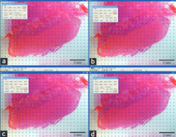
Structural changes in the rat placenta during the last third of gestation were for the first time assessed by stereology. Fischer female rats were euthanized on the day 16 or day 19 of gestation, and 35 placentas were collected. Three randomly selected placentas from each group were stereologically analyzed for the absolute volume. The proportion of the glycogenic cells and the trophoblast giant cells (TGC) in the basal part of the placenta was calculated using volume density. The absolute volume of the rat placenta on the day 16 of gestation was determined as 0.0638 cm3. The labyrinth comprised 0.0274 cm3, the basal plate 0.0271 cm3 and the decidua 0.0093 cm3. On the day 19 of gestation, the absolute volume of the placenta was 0.1627 cm3, the labyrinth occupied 0.0922 cm3, the basal plate 0.0596 cm3 and the decidua 0.0109 cm3. The volume density of trophoblast giant cells was 0.174 cm0 on the day 16 and 0.107 cm0 on the day 19 of gestation. The glycogenic cells comprised 0.379 percentage of the basal plate on the day 16 and 0.236 on the day 19 of gestation. We conclude that the absolute volume of the whole placenta and the labyrinth has increased from day 16 to the day 19 of gestation. In contrast, the volume density of glycogenic cells and trophoblast giant cells was higher on the day 16 than on the day 19 of gestation, probably due to the intensive trophoblast invasion during that time.
Citations
Downloads
References
Longo LD, Reynolds LP. Some historical aspects of understanding placental development structure and function. Int J Dev Biol 2010; 54 (2-3): 237-255.
http://dx.doi.org/10.1387/ijdb.082774ll
Serman A, Serman L. Development of placenta in a rodent – model for human placentation. Front Biosci 2011; 3: 233-239.
http://dx.doi.org/10.2741/e238
Sasaki H. Mechanisms of trophoectoderm fate specification in preimplantation mouse development. Dev Growth Differ 2010; 52: 263-273.
http://dx.doi.org/10.1111/j.1440-169X.2009.01158.x
Šerman LJ, Šerman A. Uloga glikoproteina u procesima implantacije i placentacije. Gynaecol Perinatol 2006; 15(2):82-88.
Welsh AO, Enders AC. Chorioallantoic formation in the rat: I. Luminal epithelial cell death and extracellular matrix in the mesometrial region of implantation chambers. Am J Anat 1991; 192(3):215-231.
http://dx.doi.org/10.1002/aja.1001920302
Enders AC, Blankenship TN. Comparative placental structure. Adv Drug Deliv Rev 1999; 38(1):3-15. http://dx.doi.org/10.1016/S0169-409X(99)00003-4
De Rijk EPCT, Van Esch E, Filk G. Pregnancy dating in the rat: placental morphology and maternal blood parameters. Toxicol Pathol 2002; 30(2):271-282.
http://dx.doi.org/10.1080/019262302753559614
Baczyk D, Drewlo S, Proctor L, Dunk C, Lye S, Kingdom J. Glial cell missing-1 transcription factor is required for the differentiation of the human trophoblast. Cell Death Differ 2009; 16:719-727.
http://dx.doi.org/10.1038/cdd.2009.1
Carter AM Enders AC. Comparative aspects of trophoblast development and placentation. Reprod Biol Endocrinol 2004; 2(1):46-61.
http://dx.doi.org/10.1186/1477-7827-2-46
Zybina TG, Kaufmann P, Frank HG, Freed J, Kadyrov M, Biesterfeld S. Genome multiplication of extravillious trophoblast cells in human placenta in the course of differentiation and invasion into endometrium and myometrium I. Dynamics of polyploidization. Tsitologiia 2002; 44(11):1058-1067.
Huppertz B, Burton G, Cross JC, Kingdom JCP. Placental morphology. From molecule to mother-A dedication to Peter Kaufmann-A review. Placenta 2006; 27:S3-S8.
http://dx.doi.org/10.1016/j.placenta.2006.01.007
Cross JC, Baczyk D, Dobric NM, Hemberger M, Hughes M, Simmons DG, et al. Genes, development and evolution of the placenta. Placenta 2003; 24:123-130.
http://dx.doi.org/10.1053/plac.2002.0887
Carter AM. Sources of comparative studies of placentation I. embryological collections. Placenta 2008; 29:95-98.
http://dx.doi.org/10.1016/j.placenta.2007.09.008
Coan PM, Ferguson-Smith AC, Burton GJ. Developmental dynamics of the definitive mouse placenta assessed by stereology. Biol of Reprod 2004; 70: 1806-1813.
http://dx.doi.org/10.1095/biolreprod.103.024166
Pijnenborg R, Vercruysse L. Mathias Duval on placental development in mice and rats. Placenta 2006; 27: 109-118.
http://dx.doi.org/10.1016/j.placenta.2005.01.009
Serman L, Vlahovic M, Sijan M, Bulic-Jakus F, Serman A, Sincic N, et al. The impact of 5-azacytidine on placental weight, glycoprotein pattern and proliferating cell nuclear antigen expression in rat placenta. Placenta 2007; 28: 803-811.
http://dx.doi.org/10.1016/j.placenta.2007.04.001
Mayhew TM. A stereological perspective on placental morphology in normal and complicated pregnancies. Journal of Anatomy 2009 215:77-90.
http://dx.doi.org/10.1111/j.1469-7580.2008.00994.x
Veras MM, Damaceno Rodrigues NR, Caldini EG, Maciel Ribeiro AA, Mayhew TM, Saldiva PH, et al. Particulate urban air pollution affects the functional morphology of mouse placenta. Biol Reprod 2008; 79: 578-584.
http://dx.doi.org/10.1095/biolreprod.108.069591
Geusens N, Verlohren S, Luyten C, Taube M, Hering L, Vercruysse L, et al. Endovascular trophoblast invasion, spiral artery remodelling and uteroplacental haemodynamics in a transgenic rat model of pre-eclampsia. Placenta 2008; 29 :614-623.
http://dx.doi.org/10.1016/j.placenta.2008.04.005
Lyall F. Priming and remodelling of human placental bed spiral arteries during pregnancy. Placenta 2005; 26 (Suppl A): S31-36.
http://dx.doi.org/10.1016/j.placenta.2005.02.010
Camp EA, Coker AL, Robboy SJ, Noller KL, Goodman KJ, Titus Ernstoff LT, et al. Breast cancer screening in women exposed in utero to diethylstilbestrol. J Womens Health (Larchmt) 2009; 18: 547-552.
Downloads
Additional Files
Published
How to Cite
Accepted 2014-12-03
Published 2015-01-22