Skeletal muscle myosin heavy chain expression and 3D capillary network changes in streptozotocin-induced diabetic female mice
DOI:
https://doi.org/10.17305/bb.2023.9843Keywords:
Type 1 diabetes mellitus (T1DM), skeletal muscle, myosin heavy chain (MyHC) isoforms, capillary network analysis, muscle fiber composition, three-dimensional (3D) image analysisAbstract
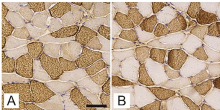
It is not well-understood how type 1 diabetes (T1DM) affects skeletal muscle histological phenotype, particularly capillarisation. This study aimed to analyze skeletal muscle myosin heavy chain (MyHC) fibre type changes and 3D capillary network characteristics in experimental T1DM mice. Female C57BL/6J-OlaHsd mice were categorized into streptozotocin (STZ)-induced diabetic (n = 12) and age-matched non-diabetic controls (n =12). The muscle fibre phenotype of the soleus, gluteus maximus, and gastrocnemius muscles were characterized based on the expression of MyHC isoforms, while capillaries of the gluteus maximus were assessed with immunofluorescence staining, confocal laser microscopy and 3D image analysis. STZ-induced diabetic mice exhibited elevated glucose levels, reduced body weight, and prolonged thermal latency, verifying the T1DM phenotype. In both T1DM and non-diabetic mice, the gluteus maximus and gastrocnemius muscles predominantly expressed fast-twitch type 2b fibers, with no significant differences noted. However, the soleus muscle in non-diabetic mice had a greater proportion of type 2a fibers and comparable type 1 fiber densities (26.2 ± 14.6% vs 21.9 ± 13.5%) relative to diabetic mice. T1DM mice showed reduced fiber diameters (P = 0.026), and the 3D capillary network analysis indicated a higher capillary length per muscle volume in the gluteus maximus of diabetic mice compared to controls (P < 0.05). Overall, T1DM induced significant changes in the skeletal muscle, including shifts in MyHC fibre types, decreased fibre diameters, and increased relative capillarisation, possibly due to muscle fibre atrophy. Our findings emphasize the superior detail provided by the 3D analytical method for characterizing skeletal muscle capillary architecture, highlighting caution in interpreting 2D data for capillary changes in T1DM.
Citations
Downloads
Downloads
Published
Issue
Section
Categories
License
Copyright (c) 2023 Nejc Umek, Luka Pušnik, Chiedozie Kenneth Ugwoke, Žiga Šink, Simon Horvat, Jiří Janáček, Erika Cvetko

This work is licensed under a Creative Commons Attribution 4.0 International License.
How to Cite
Accepted 2023-10-18
Published 2024-05-02


