Differences between inflammatory cells infiltrated into tunica intima, media, and adventitia of ascending aortic aneurysms within diabetic and hypertensive patients
DOI:
https://doi.org/10.17305/bb.2022.8565Keywords:
type 2 diabetes (DM), arterial hypertension (AH), ascending aortic aneurysm, intima, media, adventitia, inflammationAbstract
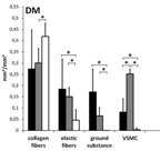
The risk factors that are the most significant for the development of most cardiovascular diseases are arterial hypertension (AH), type 2 diabetes (DM), and inflammation. However, for the development of aortic aneurysms, DM is not one of them. Our study aimed to evaluate the difference between inflammatory infiltration in three individual layers of the ascending aortic aneurysm within diabetic and hypertensive patients. Forty-five patients aged 36 to 80 were divided into a group with diabetic patients without AH (group DM, N=8) and hypertensive patients without DM (group AH, N=37). For the histological analysis, aortic aneurysms were stained with hematoxylin eosin and Movat. We used immunochemical methods to detect pro- (M1), anti-inflammatory (M2) macrophages, T-helper, T-killer cells, B cells, and plasma cells. Statistical analysis was done by independent-samples Kruskal-Wallis test adjusted by Bonferroni correction for multiple tests (P<0.05). We found no difference in the volume density of collagen, elastin, vascular smooth muscle cells (VSMC), and ground substance between groups. In the DM group, there were significantly fewer M2, T-helpers, and T-killers in the media than in the intima and the adventitia (P<0.05). There were no significant differences in the number of M1, B, and plasma cells between all three vascular layers (P<0.05). In the AH group, there were significantly fewer B and plasma cells, T-helper, T-killer cells, M1, and M2 in the media than in the intima and adventitia (P<0.05). Our results conclude that the tunica media in the aneurismal wall of the AH group retained immune privilege. In contrast, in the DM group, all three layers were immune-privileged.
Citations
Downloads
Downloads
Additional Files
Published
License
Copyright (c) 2023 Aleksandra Milutinović, Ruda Zorc-Pleskovič

This work is licensed under a Creative Commons Attribution 4.0 International License.
How to Cite
Accepted 2023-01-12
Published 2023-07-03