Role and significance of c-KIT receptor tyrosine kinase in cancer: A review

Authors

  • Emana Sheikh OMS-III, Dr. Kiran C. Patel College of Osteopathic Medicine, Nova Southeastern University, Fort Lauderdale, Florida, United States
  • Tony Tran Dr. Kiran C. Patel College of Osteopathic Medicine, Nova Southeastern University, Fort Lauderdale, Florida, United States.
  • Semir Vranic College of Medicine, QU Health, Qatar University, Doha, Qatar. https://orcid.org/0000-0001-9743-7265
  • Arkene Levy Department of Medical Education, Dr. Kiran C. Patel College of Allopathic Medicine, Nova Southeastern University, Fort Lauderdale, Florida, United States.
  • R. Daniel Bonfil Department of Medical Education, Dr. Kiran C. Patel College of Allopathic Medicine, Nova Southeastern University, Fort Lauderdale, Florida, United States.

DOI:

https://doi.org/10.17305/bjbms.2021.7399

Keywords:

c-KIT, CD117, cancer, receptor tyrosine kinases, stem cell factor receptor

Abstract

c-kit is a classical proto-oncogene that encodes a receptor tyrosine kinase (RTK) that responds to stem cell factor (SCF). C-KIT signaling is a critical regulator of cell proliferation, survival, and migration and is implicated in several physiological processes, including pigmentation, hematopoiesis and gut movement. Accumulating evidence suggests that dysregulated c-KIT function, caused by either overexpression or mutations in c-kit, promotes tumor development and progression in various human cancers. In this review, we discuss the most important structural and biological features of c-KIT, as well as insights into the activation of intracellular signaling pathways following SCF binding to this RTK. We then illustrate how different c-kit alterations are associated with specific human cancers and describe recent studies that highlight the contribution of c-KIT to cancer stemness, epithelial-mesenchymal transition and progression to metastatic disease in different experimental models. The impact of tyrosine kinase inhibitors in treating c-KIT-positive tumors and limitations due to their propensity to develop drug resistance are summarized. Finally, we appraise the potential of novel therapeutic approaches targeting c-KIT more selectively while minimizing toxicity to normal tissue.

Downloads

Download data is not yet available.
Role and significance of c-KIT receptor tyrosine kinase in cancer: A review

Downloads

Additional Files

Published

2022-09-16

How to Cite

1.
Sheikh E, Tran T, Vranic S, Levy A, Bonfil RD. Role and significance of c-KIT receptor tyrosine kinase in cancer: A review. Biomol Biomed [Internet]. 2022Sep.16 [cited 2023Nov.29];22(5):683-98. Available from: https://bjbms.org/ojs/index.php/bjbms/article/view/7399

Issue

Section

Reviews

INTRODUCTION

About two-thirds of the 90 tyrosine kinase (TK) genes described in the human genome encode for receptor tyrosine kinases (RTKs) [1]. These cell-surface receptors transduce a response on binding to a ligand and are defined by an extracellular (EC) ligand-binding domain, a single transmembrane (TM) region, a juxtamembrane (JM) region, a cytoplasmic portion with a conserved protein TK domain, and a flexible carboxy (C)-terminal tail [2,3]. RTKs are ubiquitously spread in multicellular animals, from the oldest metazoan phylum existing today (Porifera) to Chordata [4]. In humans, the 58 RTKs described so far are classified into 20 subfamilies or classes based on the structure of their amino (N)-terminal ligand binding ectodomains, which consist of one or more defined motifs including cysteine-rich regions, fibronectin type III-like domains, immunoglobulin (Ig)-like domains, kringle-like domains, epidermal growth factor-like domains, cadherin-like domains, discoidin-like domains, and leucine-rich regions [1,5]. Among the different classes of human RTKs described to date, Class III RTKs, which are characterized by the presence of five Ig-like EC domains, include platelet-derived growth factor α and β receptors (PDGFR α/β), colony-stimulating factor 1 receptor, fms-like RTK 3, and c-KIT [6,7]. These RTKs play a pivotal role in several aspects of normal cell physiology, and different mutations that affect them can cause aberrant downstream signaling that is often linked to many disorders, including cancer [8,9].

STRUCTURE AND BIOLOGICAL FUNCTIONS OF C-KIT

The proto-oncogene c-kit, mapped to chromosome 4q11-12 in humans [2] and chromosome 5 (W locus) in mice [10], was discovered in 1986 as the cellular homolog of the transforming viral oncogene v-kit in the Hardy-Zuckerman 4 feline sarcoma virus [11]. Wild-type c-kit encodes for a 145 kDa, 976 amino acid type IIIa RTK protein known as c-KIT, which is often referred to as CD117 or stem cell factor (SCF) receptor due to its association with its ligand SCF [12]. The c-KIT protein resides in the cell membrane and is comprised of EC, TM, and intracellular (IC) regions (Figure 1). Like all class III RTKs, the EC portion of c-KIT comprises five Ig-like domains (D1-D5). The first three domains are essential for c-KIT binding to SCF, whereas D4 and D5 are involved in dimerizing adjacent c-KIT monomers [13,14]. The EC region is followed by a single spanning TM helix that connects with the IC domain, including a JM domain coupled to a TK domain and a C-terminal tail region. The JM domain is essential for c-KIT receptor control and modulation, particularly in the relay of IC downstream signaling [15]. The TK domain is split into the proximal amino-terminal lobe (N-lobe) TK1 with an ATP-binding region, and a distal carboxy-terminal lobe (C-lobe) TK2 with a phosphotransferase domain [16] (Figure 1).

FIGURE 1: Structural organization of the human c-KIT receptor. In its inactivated state, c-KIT is present as a monomer that comprises extracellular (EC), transmembrane (TM) and intracellular (IC) domains. The outer immunoglobulin-like (Ig-like) domains D1 to D3 are key components for binding to stem cell factor (SCF), whereas D4 and D5 are essential for homotypic contacts needed for KIT dimerization. The IC domain contains a juxtamembrane (JM) domain, a tyrosine kinase (TK) domain and a flexible carboxy-terminal (C-terminal) tail. The JM domain contributes to the relay of IC downstream signaling. The TK domain is further divided into the amino-terminal TK1 (N-lobe) domain, which houses an ATP-binding region, and the carboxy-terminal TK2 (C-lobe) domain, which encompasses a phosphotransferase region and activation loop.

Different c-KIT isoforms generated by alternative mRNA splicing have been described, including two that differ by the presence or absence of the tetrapeptide sequence glycine-asparagine-asparagine-lysine (GNNK) in the EC domain [17-19]. Although both c-KIT isoforms have binding affinity to SCF, the GNNK-negative isoform leads to faster phosphorylation of the receptor, a more robust downstream signaling, and higher tumorigenic potential in mice [20-23]. Another c-KIT isoform results from losing one of the two serine residues in the kinase insert (KI) domain [17]. In contrast, a fourth isoform is caused by a shorter transcript of c-Kit that encodes a truncated c-KIT without kinase activity and only contains TK2 and the C-terminal tail region [24].

c-KIT is expressed by various cells in the body, and signaling pathways stimulated by its activation by SCF under physiologic conditions are implicated in regulating cellular processes such as cell proliferation, survival and migration [13]. In the normal bone marrow, c-KIT is expressed by hematopoietic stem cells, playing an important role in self-renewal and differentiation into various blood cells (reviewed in [25]). Indeed, homozygous white-spotted (W) loss-of-function mutations in the c-Kit gene (c-KitW/W) in mice have shown to cause lethal anemia caused by hematopoietic stem cell defects [26,27]. c-KIT expression is gradually lost during hematopoietic differentiation and only retained or increased in mast cells, natural killer (NK) cells and dendritic cells (DCs), suggesting an essential function in inflammation and immunity [28,29]. Moreover, different studies have shown that CD117/c-KIT is not only expressed by bone marrow-derived stem cells, but also by those found in other organs in adults, such as prostate [30], liver [31] and heart [32], suggesting that SCF/c-KIT signaling pathways may contribute to stemness in some organs. Furthermore, c-KIT has been linked to many different biological processes in other cell types. For instance, c-KIT signaling has been shown to regulate oogenesis, folliculogenesis and spermatogenesis, exerting critical functions in female and male fertility [33,34].

c-KIT is also critical to the proliferation, survival and migration of melanocytes from the neural crest to the dermis [35]. Loss-of-function mutations in c-kit can cause piebaldism, an autosomal dominant disorder characterized by congenital absence of melanocytes in patches of skin and hair, similar to the “dominant white spotting” observed in mice with mutations in the same gene [36,37]. In individuals with the piebald trait, constipation is often seen because of the loss of interstitial cells of Cajal (ICC), which are c-KIT-positive cells that control gut peristalsis [36]. The role of c-KIT in ICC development is supported by studies in mice with loss-of-function mutations in c-Kit that present a constipation phenotype [38,39].

ACTIVATION AND DOWNSTREAM SIGNALING OF C-KIT

Under physiological conditions and when it is not bound to SCF, c-KIT resides in the cell membrane as a monomer. In this resting state, c-KIT is cis-autoinhibited by the JM domain that inserts between the TK1 (N-lobe) and TK2 (C-lobe) domains. This leads to a static configuration that sterically blocks the “activation loop” that resides in the catalytic cleft between the lobes from assuming an extended and active conformation (“JM autoinhibition”) [15,40,41] (Figure 2A). The binding of dimeric SCF to D1-D3 regions bridges two adjacent c-KIT molecules together and leads to a D4 and D5 reorientation that results in c-KIT homodimerization [42,43]. This conformational change leads to trans-autophosphorylation of selected tyrosine residues (Y) events that appear to occur in a specific order. The initial autophosphorylation occurs in tyrosine residues in the JM domain (primarily Y568 and Y570), resulting in its displacement from the N-lobe and swinging away of the loops, allowing access to ATP and release of ADP from the active site [5,13,15,16]. Subsequent transphosphorylation occurs in the activation loop (Y823) [13,15,16] (Figure 2B). Full activation of c-KIT occurs when additional tyrosine residues in the KI region (Y703, 721, and 730) and the C-terminal tail (Y900 and Y936) are phosphorylated [13,15,16] (Figure 2C).

FIGURE 2: Schematic representation of c-KIT activation and downstream signaling. (A) Here, two adjacent monomeric c-KIT molecules are cis-autoinhibited by the juxtamembrane (JM) domain that inserts between the tyrosine kinase (TK) 1 and 2 domains, leading to a static configuration that sterically blocks the activation loop (AL) residing in the catalytic cleft between the lobes; (B) c-KIT is activated on binding of dimeric stem cell factor (SCF) to immunoglobulin-like (Ig-like) domains D1 to D3, which induces receptor reorientation and homotypic interaction between adjacent D4 and D5 domains. Transphosphorylation of tyrosine residues in the JM domain enables its dissociation from the TK1 domain. TK1 and TK2 domains are displaced away, allowing access to the catalytic cleft for ATP binding and release of ADP from the binding site for a second transphosphorylation of tyrosine residues in the AL; (C) Further phosphorylation of tyrosine residues in the kinase insert region and C-terminal tail creates docking sites for several substrates, leading to downstream signaling through the mitogen-activated protein kinase/extracellular signal-regulated kinase (MAPK/ERK), phosphatidylinositol 3-kinase/protein kinase B (PI3K/AKT), phospholipase C-γ (PLC-γ), Janus kinase/signal transducer and activator of transcription (JAK/STAT), and Src kinase pathways.

Many of the tyrosine residues mentioned above serve as substrate docking sites after transphosphorylation, activating downstream transduction pathways that lead to various cellular responses (Figure 2C). It is important to note that the transduced signaling pathways and their consequential effects are dependent on the specific tyrosine residue phosphorylated. Here, we will provide a simplified explanation of signaling pathways downstream of c-KIT that, rather than being just linear and independent, are now known to be much more complex and occasionally connected with other downstream signaling molecules activated by other receptors.

Signaling cascades activated downstream of c-KIT include mitogen-activated protein kinase/EC signal-regulated kinase (MAPK/ERK), phosphatidylinositol 3-kinase/protein kinase B (PI3K/AKT), C-γ (PLC-γ), Janus kinase/signal transducer and activator of transcription (JAK/STAT), and Src kinase pathways [44]. On phosphorylation, Y703 and Y936 bind the SH2 domain of the adaptor protein growth factor receptor-bound protein 2 (Grb2), ending in the activation of the MAPK/ERK pathway, which plays essential roles in the regulation of gene transcription and cell proliferation [45,46]. Conversely, phosphorylation of c-KIT at Y721 triggers the PI3K/AKT pathway that promotes cell survival and evasion of apoptosis, either through direct binding of the p85 subunit of PI3K or indirectly through binding of PI3K to the scaffolding protein Grb2-associated binding protein (Gab2) and Grb2 [47-49]. The PI3K/AKT pathway can also be activated by phosphorylated Tyr900 in the C-lobe through binding to the adaptor protein Crk [41]. The PLC-γ pathway, which promotes cellular proliferation and suppresses apoptosis through the actions of diacylglycerol and inositol 1.4,5-triphosphate, can be triggered when PLC-γ interacts with phosphorylated Y730 [13,50]. In addition, activation of the Src family of tyrosine kinases (SFK) has been reported to occur through the interaction of its SH2 domain with phosphorylated Y568, Y570, and Y936, stimulating cell proliferation and survival through Akt phosphorylation and, presumably, cell migration through phosphorylation of focal adhesion kinase [46,51]. Moreover, SFK and PI3K/AKT participate in the activation of the JAK/STAT pathway [46,52], suggesting that phosphorylation of some c-KIT tyrosine residues leads to activation and translocation of STAT proteins into the nucleus, where they act on target gene promoters [53,54].

Different regulatory mechanisms function as negative-feedback loops to ensure tight control of signaling output once the c-KIT receptor is activated [55]. The main mechanisms of attenuation of c-KIT signaling include (1) c-KIT ubiquitination and internalization, (2) dephosphorylation, and (3) PKC-dependent serine phosphorylation. In the first of these mechanisms, activated c-KIT is transported from the cell surface to the interior of the cell through clathrin-mediated endocytosis [56]. E3 ubiquitin-protein ligase c-Cbl (named after Casitas B-lineage Lymphoma) binds directly to activated c-KIT receptors through Y568 and Y936 or indirectly through Grb2 to Y703 and Y936, or via the p85 subunit of PI3K [13,57,58]. Binding of another E3 ubiquitin ligase complex containing suppressor of cytokine signaling (SOCS) 1 and 6 isoforms to Y568 in activated c-KIT has also been described [59,60]. The internalized c-KIT is then targeted for lysosomal and proteasomal degradation [61,62]. Furthermore, attenuation of c-KIT signaling can occur by the action of phosphatases such as Src homology region 2 domain-containing phosphatase-1, which has been shown to associate with activated c-KIT causing its dephosphorylation [63,64]. Finally, increased PKC activation downstream from c-KIT can lead to negative feedback regulation of the receptor by phosphorylating Ser741 and Ser746 residues in the KI domain [65,66]. Moreover, PKC activation has also been shown to cause shedding of the EC domain of c-KIT, making it unresponsive to SCF stimulation [67].

C-KIT AND CANCER

Genomic profiling of nearly 19,000 de-identified samples has shown c-kit alterations in 2.86% of 59 major cancer types studied, with some of them presenting very frequent and clinically actionable mutations [68], such as gastrointestinal stromal tumor (GIST) in about 80-85% of cases [69]. Although most c-kit alterations associated with cancer involve “gain-of-function” mutations that lead to constitutive activation of c-KIT in an SCF-independent manner, others entail amplification/overexpression or “loss-of-function” mutations [70].

Gain-of-function c-kit mutations have been found to represent oncogenic driver events in the development of a wide variety of cancers/proliferative diseases, including GISTs [71,72], some subtypes of melanomas [73], mastocytosis [74], acute myeloid leukemia (AML) [75], and seminomas [76].

In GIST, c-KIT expression is detected immunohistochemically in more than 95% of the cases. It has become an important diagnostic marker when used together with morphologic features displayed by these tumors [77,78]. In addition, 85%–90% of adult GISTs bear c-kit and PDGFRA gene gain-of-function mutations that are mutually exclusive [69] and seem unrelated to c-KIT expression, as they can be found in a proportion of GISTs that are immunohistochemically negative for c-KIT [77]. Mutations in c-kit, which in GISTs are more common than those in PDGFRA, most frequently involve the exon 11 that codes for the JM region, disrupting its autoinhibitory function and leading to constitutive activation of c-KIT [71,79]. Most of the mutations in exon 11 are caused by deletions and clusters between codons 550 and 560, which represents one of the hot spots within the c-kit gene [13,69]. Less common mutations occur in exon 9 that encodes the EC region of c-KIT, mainly involving an internal tandem duplication of Ala502-Tyr503 [71,80,81] that would mimic the conformational change that occurs when c-KIT dimerizes after binding its cognate ligand SCF [82]. Mutations also occur in exons 13 (encoding the ATP-binding region of c-KIT) and 17 (encoding the activation loop of the kinase), but are rare, with a combined frequency of 1-2% among all GISTs [81,83,84].

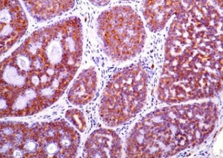
In about 80% of melanomas, the main oncogenic drivers involve mutations in B-Raf proto-oncogene serine/threonine kinase (BRAF) and neuroblastoma rat sarcoma viral oncogene homolog (NRAS) (reviewed in [85]). However, c-kit mutations, which are typically mutually exclusive of BRAF and NRAS mutations, are identified in around 3% of melanomas, particularly those derived from acral surfaces (soles, palms, and nail beds) (36%), mucosa (39%), and chronically sun-damaged sites (28%) (reviewed in Reddy et al. [85], Pham et al. [86]). About 70% of mutations affecting c-kit in melanomas are constitutive activating mutations, including L576P (lysine-to-proline mutation at codon 576) in exon 11 and K642E (methionine-to-glutamic mutation at codon 642) in exon 13 [86]. Besides gene alterations, immunohistochemical studies have revealed overexpression of c-KIT in some melanoma variants, particularly among ocular melanomas (36-91%) [87-89]. In these cases, overexpression of c-KIT seems unrelated to c-kit mutations in exons 11, 13, 17, or 18 [88,89]. Moreover, c-KIT overexpression was associated with a worse outcome in patients with choroidal and ciliary body melanoma [90]. Figure 3A and B show representative immunohistochemical staining of a c-KIT-positive uveal melanoma.

FIGURE 3: Examples of different cancer types expressing c-KIT. (A and B): Uveal melanoma morphology (A) with a strong and diffuse c-KIT expression; (C and D): Ovarian dysgerminoma (C) with C-KIT expression in cancer cells; tumor-infiltrating lymphocytes were negative (D); Renal oncocytoma (E) and mammary adenoid cystic carcinoma (F) exhibiting C-KIT positivity. All cases were stained immunohistochemically using polyclonal A-4502 antibody (DAKO Agilent). The images were taken at ×20 magnification except for C and D (×10 magnification).

Systemic mastocytosis is a rare myeloproliferative neoplasm in which malignant mast cells infiltrate bone marrow and other extracutaneous tissues such as liver, spleen, and peripheral blood. More than 90% of adult patients with this disease present a gain-of-function mutation in exon 17 within the c-kit gene, particularly KIT D816V, a missense mutation in which aspartic acid is substituted by valine in codon 816 [13,91]. Through kinase assays, it has been shown that the D816V mutant can autoactivate 586-fold faster than native c-KIT [79], which explains the adverse prognostic impact of the c-kit mutation in this hot spot in patients with systemic mastocytosis [91]. The D816V mutation is also present in children with systemic mastocytosis but with a lower frequency (42%). In contrast, other mutations occur in other locations, often in exons 8 and 9 (44%) that encode the fifth EC Ig-like domain [74], thus promoting a conformational change that enables dimerization and activation of c-KIT by lower physiologic SCF levels than normally needed [16].

c-KIT expression is seen in myeloblasts in 65-90% of AML patients [92,93] and, in some cases, co-expressed with SCF, suggesting a potential autocrine activation [13,92]. Furthermore, c-kit mutations are found in AML patients, predominantly associated with core-binding factor leukemias, an AML variant that involves chromosomal abnormalities t(8;21)(q22;q22) or inv(16)(p13q22)/t(16;16)(p13;q22) [94]. Most of these constitutive activating mutations mainly reside in exon 8 (as in-frame insertions or deletions that affect an EC domain involved in c-KIT dimerization) or exon 17 (as missense mutations that affect the activation loop in the c-KIT TK domain) [94].

Although an overall somatic mutation rate of about 8% has been reported in both seminoma and non-seminoma testicular germ cell tumors, the incidence of c-kit mutation is ten-fold higher (20-25%) in the former than in the latter [95,96]. The most common c-kit alteration in seminomas involves activating mutations in exon 17, mainly D816X (where X is either valine [V] or histidine [H]) [13,97]. Similar c-kit mutations have been reported in dysgerminomas (the ovarian counterpart of seminomas) [98,99], along with c-kit amplification associated with c-KIT protein overexpression evident through immunohistochemistry (IHC) (Figure 3C and D) [99]. Moreover, high expression of c-KIT in patients with primary ovarian high-grade serous carcinoma has been shown to be associated with shorter disease-free survival and peritoneal metastasis [100]. No correlation was found between c-kit mutations and c-KIT protein expression [99], which is detected in 78%–100% of ovarian dysgerminomas [101].

In addition to gain-of-function mutations described above for some cancers, different studies have shown overexpression of c-KIT in cancer cells that, in their normal cell counterparts, show very little or undetectable c-KIT expression when mainly assessed by IHC. For example, a 7-fold increase in c-kit mRNA expression relating to normal renal tissue has been reported in renal oncocytoma and chromophobe renal cell carcinoma (RCC) [102]. Moreover, IHC analysis performed in tissue microarrays (TMAs), including 226 renal tumors, revealed a strong c-KIT immunoreactivity in more than 85% of chromophobe RCCs and oncocytomas. In contrast, c-KIT expression was infrequently observed or undetectable in other renal tumors assessed [102,103]. IHC studies revealed c-KIT expression in 100% of cystic renal oncocytomas (Figure 3E) [104]. c-KIT overexpression in chromophobe RCC and renal oncocytoma was not associated with c-kit mutations [105].

Normal breast ducts and acini, but not myoepithelial and stromal cells, show some expression of c-KIT, which has been reported to be lost in most breast cancers [106-108]. This loss of c-KIT expression has been a potential determinant of malignant breast transformation due to c-kit gene promoter DNA hypermethylation [109]. Although a low percentage of breast carcinomas express c-KIT, if any, 20-42% of triple-­negative breast cancers, which lack expression of estrogen receptor, progesterone receptor, and HER-2/neu and have a significantly higher probability of relapse and poorer overall survival when compared with other breast cancer types, do express it [110-112]. Another type of breast cancer in which the expression of c-KIT is frequently seen is adenoid cystic carcinoma (ACC) of the breast that, although relatively clinically indolent, can be confounded with infiltrating duct carcinomas (particularly with tubular and cribriform carcinomas of the breast). In this context, IHC assessment of c-KIT is a valuable diagnostic tool since its expression is found in more than 90% of mammary ACC but not in other carcinomas with overlapping histologic features (Figure 3F) [113-115].

Although overexpression of c-KIT - rather than mutations in its gene - has been reported in a high percentage of small cell lung cancer (SCLC) patients by different groups [116-120], its prognostic relevance remains debatable due to conflicting findings that may be related to the type of tumor specimens used (biopsy or surgical samples), cancer stages, and other variables that still need to be scrutinized.

Expression of c-KIT and SCF has been reported in patient-derived immortalized colorectal cancer cell lines [121] and in premalignant and malignant colonic lesions, where c-KIT and SCF co-expression has been associated with a worse clinical outcome [122]. In a more recent study using a TMA comprising 137 patient-derived colon tumors and 179 associated serially passaged xenografts, it was found that c-KIT is expressed in approximately 50% of colorectal cancer tissues [123], in agreement with data collected from The Cancer Genome Atlas [124].

Several studies have assessed c-KIT expression in human prostate cancer (PCa) cell lines and biopsies taken from patients, though with some divergent results that may be due to the use of antibodies that have been discontinued some years ago and thus cannot be further employed for reproducibility analyses [125,126]. Studies carried out by our lab (RDB) using benign prostatic hyperplasia, primary tumors, and bone metastatic PCa specimens have shown uniform levels of SCF and a trend of increasing c-KIT expression that parallels disease aggressiveness [127]. These findings are in agreement with other studies that also revealed significantly increased expression of c-KIT in high-grade (Gleason score [GS] 8 or higher, or clinical Stage 2) compared with low-grade (GS 6-7, or clinical Stage 2) prostate tumors [128]. Despite this, we observed that most human PCa cell lines grown in vitro express c-kit at the gene level, though c-KIT immunoblotting only detects low or null protein levels [127], as shown similarly by others [129]. Using experimental models of bone metastasis, we observed de novo expression of c-KIT in intraosseous tumors generated by otherwise c-KIT-negative PCa cell lines, suggesting an induction of c-KIT expression in PCa cells by the bone microenvironment, which was confirmed by co-culture studies of PCa cells and bone marrow-derived cells [127,130]. Furthermore, we found that inhibition of bone-induced c-kit expression in PCa cells transduced with lentiviral short hairpin RNA could significantly reduce intraosseous tumor incidence and growth, suggesting a crucial role of this RTK in PCa bone metastasis [130].

IMPLICATIONS OF C-KIT IN CANCER DEVELOPMENT AND PROGRESSION

A complex interplay of numerous biological processes contributes to the development and progression of cancer. In addition to studies reporting associations between specific gain-of-function or loss-of-function mutations in c-kit, induction of de novo expression or overexpression of c-KIT and clinical outcome in cancer patients (summarized above), research by many groups has revealed that c-KIT plays crucial roles in the regulation of many mechanisms leading to tumor formation and cancer progression in carcinomas. Below, we will describe some of these studies.

Cancer stemness, which refers to the cancer stem cell (CSC) phenotype, is characterized by the ability of a subpopulation of cancer cells to self-renew, differentiate into defined progenies, initiate tumor growth, and drive metastasis, recurrence, and resistance to therapies [131,132]. C-KIT has been proposed to regulate stemness in different cancers. Studies in ovarian cancer cells have related c-KIT expression to cancer stemness [133-136]. Accumulating evidence also indicates a role for c-KIT in colon cancer stemness, as supported by studies employing spheroid cultures derived from colon cancer patients’ tumor cells grown in serum-free and non-adherent plates, a technique commonly used to investigate CSCs [137]. These studies have shown that the release of SCF by more differentiated colon tumor cells modulates the growth of c-KIT-expressing CSC-like colon tumor cells [138], suggesting the existence of a paracrine system by which SCF can stimulate CSC-like cells found in the colonospheres. Furthermore, it was recently demonstrated that c-KIT stimulates CSC properties in colorectal cancer cells, including CD44 expression and other stem cell markers [139]. Studies on non-small lung cancer have also related c-KIT to cancer stemness, based on findings that revealed a reduction of CSCs through targeting the SCF-c-KIT autocrine signaling loop [140] and inhibition of c-kit with specific shRNA and inhibitors [141]. Moreover, in a recent study in which human PCa cell lines were separated into CD117 (c-KIT)-positive and CD117-negative cells, Kerr’s group demonstrated that in some instances c-KIT promoted sphere formation and increased the expression of specific stemness markers [142]. Similarly, we have observed that ectopic expression of c-KIT in PC3 cells followed by exposure to its ligand SCF increases the number of prostaspheres formed in selective serum-free medium and non-adherent plate conditions (Figure 4).

FIGURE 4: c-KIT expression and sphere formation in prostate cancer cells. (A) Representative image of prostaspheres formed by PC3 cells 6 days after transfection with c-kit and exposure to SCF under non-adherent 3D culture conditions (selective serum-free medium and non-adherent plates). Note that control PC3 cells transfected with the empty vector (EV) show little homotypic cell aggregation. Scale bars, 50 μm; (B) Quantitation of sphere formation by c-KIT-expressing and EV-expressing PC3 cells 6 days after plating at different numbers in non-adherent conditions. Data are expressed as the mean ± SE number of spheres larger than 50 μm per ten 100× microscopic fields. *p=0.05; **p<0.005 (Student’s test).

To acquire migratory and invasive capacities, carcinoma cells must detach from adjacent epithelial cells and adopt a mesenchymal phenotype – the epithelial-mesenchymal transition (EMT), which plays a critical role in their aggressiveness and metastatic potential [143,144]. Regulators of EMT comprise different transcription factors such as Snail (including Snail and Slug, also called SNAI1 and SNAI2, respectively) and ZEB (Zeb1 and Zeb2), which can repress the expression of genes encoding E-cadherin and cytokeratins (associated with epithelial phenotype) and upregulate others encoding proteins linked to the mesenchymal phenotype (e.g., N-cadherin, vimentin, and fibronectin) [145-148]. Different studies have shown that the expression of EMT transcription factors is also increased in CSCs [147,149,150], and a growing body of evidence supports the view that circulating tumor cells (CTCs) can arise from tumor cells that have gone through EMT [151-154]. Acquisition of EMT properties and enhanced invasiveness and CSC traits has been found in salivary ACC cell lines after ectopic expression of c-KIT [155]. The association between KIT and EMT is also supported by immunohistochemical studies performed in a TMA comprising 150 specimens of thymic epithelial tumors, where the expression rate of c-KIT was found to be significantly higher in thymic carcinomas than in thymomas, which in most of the cases behave in a benign fashion and are noninvasive. In these studies, c-KIT expression positively correlated with EMT markers N-cadherin, Twist, and Snail and negatively with E-cadherin, suggesting that the immunohistochemical analysis of those proteins could be important to distinguish between thymic cancer and thymoma [156]. The association between c-KIT (CD117) and EMT is also supported by studies in ovarian cancer cells, where a reduction of CD117+ and CD44+ subpopulation of ovarian CSCs by metformin at low dose led to a significant decrease of Snail2, Twist, and vimentin related to mesenchymal traits, and an increase in expression of the epithelial marker E-cadherin [157]. In line with these findings, another group demonstrated that CD117+ subpopulations of human PCa cell lines present a significant increase in vimentin expression and in vitro migratory ability than CD117− subpopulations of the same cell lines [142]. In concordance with these results, we found that ectopic expression of c-KIT supports the in vitro migration and invasion of two different PCa cell lines along with BRCA2 downregulation, which may play a role in the process as suggested by gene rescue experiments [130]. We posit that the stimulation of the migratory and invasive abilities induced by c-KIT in these PCa cells would be mediated by an EMT-like phenomenon, as we observed a change in morphology toward a more mesenchymal phenotype, an increase in expression of Snail1, Slug, Zeb1, and vimentin, and a decrease in E-cadherin expression in c-kit-transfected PCa cell lines as compared to the same PCa cells transfected with the empty vector (EV) (c-kit-negative control cells) (Figure 5).

FIGURE 5: c-KIT expression and EMT-like phenomenon in prostate cancer. (A) PC3 cells stably transfected with c-kit show a fibroblast-like phenotype, while control PC3 cells transfected with the empty vector (EV) display the typical epithelial phenotype. Scale bars, 50 μm; (B) Western blots for c-KIT, epithelial-mesenchymal transition (EMT) markers, and EMT-related transcription factors are shown for PC3 and C4-2B PCa cells stably transfected with c-kit or EV and incubated with SCF (100 ng/mL for 60 min). Glyceraldehyde 3-phosphate dehydrogenase (GAPDH) served as a loading control; (C) Gene expression of Snail, Slug, and Zeb1 is higher in c-KIT-expressing PC3 and C4-2B cells relative to EV-transfected control cells (RT-qPCR; p<0.001, Student’s test). Gene expression was normalized to GAPDH. Values are mean ± SE for triplicate samples.

In addition to the contributory role exerted by cancer cell-intrinsic expression and activation of c-KIT in tumor development and progression, several lines of evidence suggest a key role for SCF-c-KIT signaling occurring in the tumor microenvironment. Perhaps the best example is that of mast cell infiltrates associated with tumors. Indeed, different studies in mice have demonstrated that high levels of c-KIT on mast cells and their presence in the tumor microenvironment promote angiogenesis, leading to increased tumor growth and metastasis [158-160]. Furthermore, additional research indicates that c-KIT and mast cells modulate the development, recruitment, and immunosuppressive effects of myeloid-derived suppressor cells in tumors [161,162].

TARGETED THERAPY OF C-KIT-POSITIVE TUMORS

As previously outlined, various cancers present an aberrant activation of c-KIT kinase, caused either by overexpression or mutations in c-kit. Most of the 500 c-kit mutations identified so far in human cancer (Sanger Institute Catalogue of Somatic Mutations in Cancer [163]are passenger rather than driver mutations. To target and inhibit dysregulated c-KIT, two main approaches have been considered: small molecule inhibitors and monoclonal antibodies (mAbs). Among small molecule inhibitors, the first one developed was imatinib mesylate (Gleevec®), which was originally found to inhibit the TK activity of the chimeric BCR-ABL fusion oncoprotein resulting from the translocation t(9;22) in chronic myelogenous leukemia, and was approved for the treatment of this hematologic cancer in 2001 [164]. Serendipitously, imatinib was also found to inhibit the autophosphorylation and activation of some RTKs, such as c-KIT and PDGFR, and was approved as standard first-line treatment for metastatic GIST. It is also used in the adjuvant setting for patients with GISTs who may have potential curative treatment by surgery and for the treatment of adult patients following surgical removal of CD117-positive GISTs (reviewed in Kelly et al. [165]). Although imatinib can traverse the cell membrane and bind to JM and cytoplasmic enzymatic domains due to its small size (Molecular Weight: 493.6), its therapeutic effect is highly dependent on the mutation involved [70]. For instance, in GIST patients whose tumors harbor gain-of-function point mutations in the exon 11 JM domain of c-kit, found in 75-80% of the cases, imatinib provides a robust initial clinical response [71]. However, in almost 90% of these patients, there is a relapse of the disease within 20-24 months [166-169], which is due to secondary mutations in c-kit that usually cluster in exons 13/14 (the ATP-binding pocket) and 17 and 18 (the activation loop) of the kinase domain, preventing optimal binding of imatinib and restoring c-KIT signaling in the presence of the inhibitor (reviewed in Serrano et al. [169]). This has led to the approval of other TK inhibitors, such as sunitinib and regorafenib, with activity against secondary c-kit mutations [169]. Among these, there are agents such as sunitinib, which elicits longer progression-free survival and overall survival in patients that harbor exon 13 or 14 secondary c-kit mutations compared to those with exon 17 or 18 secondary c-kit mutations [167,168], and regorafenib, which has equal efficacy in tumors with secondary exon 13/14 or exon 17/18 mutations, or combinations thereof [170]. Ripretinib, a novel type II switch control kinase inhibitor, is a broad-spectrum inhibitor of secondary drug resistance mutations, including activation loop mutations targeted by type I inhibitors [171]. In addition, because PDGFRA-mutant GIST accounts for up to 10% of GISTs that exhibits primary resistance to imatinib and sunitinib therapy [165], other agents that selectively target PDGFRa D842V mutant advanced GISTs are of immense clinical value. Among them, we find avapritinib, an inhibitor of c-KIT and PDGFRA activation loop mutants that has been approved by the Food and Drug Administration (FDA) for GISTs that harbor PDGFRA exon 18 D842V mutations [167], whereas dasatinib, an oral inhibitor of c-KIT, produced a positive response in one patient with PDGFRA D842V-mutant GIST in a Phase II trial, and is currently used off label for this molecular subtype [172].

The clinical experience of imatinib in GIST led to studies to explore the potential therapeutic value of this TK inhibitor in systemic mastocytosis. In adult patients affected with the disease, the activating D816V c-kit mutation is found in about 90% of the cases and is responsible for primary resistance to imatinib. In contrast, in the remaining patients (with the absence of D816V c-kit mutation or unknown c-kit mutational status), a clinical response to imatinib was found, leading to its approval as a treatment by the FDA in 2006 [173-175]. This clearly demonstrates the relevance of identifying specific c-kit mutations to select patients for more adequate treatments.

To overcome the resistance developed in certain wild-type or mutant c-KIT-positive cancers treated with TK inhibitors such as imatinib, it has been proposed the use of mAbs to target and inhibit dysregulated c-KIT. Although unlike small molecule inhibitors, antibodies can only recognize EC epitopes of c-KIT, this may represent a potential therapeutic advantage due to their specific binding to both mutant and wild-type c-KIT receptors (recall that most c-kit mutations localize to JM or IC domains of the receptor). Using KIT-expressing NIH 3T3 and Ba/F3 cell lines, Shi et al. evaluated the feasibility of targeting oncogenic c-kit mutations using anti-D4 mAbs that obstruct homotypic D4 or D5 contact formation [176]. Oncogenic c-kit mutations were divided into two classes. Class I mutants include D5 point mutations D419A and N505I, deletion of Y418 D419, and duplication of A502Y503, which exhibit surface expression of constitutively activated TK activities. In contrast, Class II mutants, including the D5 T417ID418-419 mutation and the IC V560D and D816V point mutants, have constitutively activated TK activity with low or low negligible surface expression [176]. Anti-D4 mAbs abrogated oncogenic c-KIT signaling in mutations localized in D5, including all Class I mutants and the Class II T417ID418-419 mutation. Based on these findings, the authors proposed differential pharmacological treatment regimens for cancer patients depending on the c-kit mutations present in their tumors [176].

Moreover, antibody-drug conjugates (ADCs) can also be designed by conjugating different drugs to mAbs to deliver a potent cytotoxic payload to cancer cells while minimizing toxicity to normal tissue [177]. Studies with LOP628 [178] and NN2101-DM1 [179], two humanized anti-KIT antibodies conjugated to the tubulin polymerization inhibitor emtansine (DM1) [177], have been reported. Both ADCs showed strong in vitro antiproliferative activity on several c-KIT-positive human tumor cell lines representing GIST, AML, SCLC, and systemic mastocytosis regardless of their c-kit mutational status, and in vivo antitumor responses in imatinib-sensitive and -refractory GIST and systemic mastocytosis xenograft models, as well as in SCLC and AML models [178,179]. In both cases, the ADCs bind to c-KIT on the surface of the cancer cells, forming a complex that is then internalized and rapidly trafficked to the lysosome, releasing DM1 in the cytoplasm, arresting the cell cycle by inhibiting microtubule polymerization and leading to apoptosis of cancer cells models [178,179]. Despite the promising preclinical results obtained with LOP628, rapid hypersensitivity reactions were observed in some patients treated with this ADC in a Phase I clinical trial experienced, which led to a termination of the trial [180]. This unexpected outcome is likely caused by mast degranulation resulting from a high affinity binding of the Fc region of LOP628 to the Fc-gamma receptor on mast cells and an SCF-mediated c-KIT activation that is not inhibited by LOP628 (recall that c-KIT is expressed by mast cells) [180]. Although clinical studies are still needed to define the safety profile of NN2101-DM1, it has been demonstrated that the NN2101 antibody has decreased binding affinity to Fc receptors and an inhibitory action on SCF-dependent c-KIT activation [181], which might prevent hypersensitivity reactions such those observed with LOP628 (studies in patients are necessary to examine this hypothesis). Moreover, in vivo and in vitro studies revealed synergistic inhibitory effects on some cancer cells when treated with NN2101-DM1 and imatinib or carboplatin/etoposide [179]. This suggests that the use of combination therapies involving novel anti-KIT ADCs in conjunction with standard chemotherapeutic agents, TK inhibitors, or other targeted agents, should be considered a strategy to enhance the efficacy of anti-KIT ADCs used as a monotherapy for different cancer types.

CONCLUSIONS

Knowledge of the contribution of SCF and c-KIT to different physiological mechanisms has increased dramatically during the last decades. Furthermore, accumulating evidence suggests that activating mutations or amplification/overexpression of c-kit contribute to the development and progression of many human malignancies, as supported by gene and protein profiling of clinical specimens and numerous in vitro and in vivo studies at elucidating the role played by c-KIT in cancer. Following the great success of imatinib in treating GISTs, other broad TK inhibitors have been approved to overcome the resistance acquired by certain c-KIT-positive tumors through secondary mutations occurring in c-kit. The identification of specific c-kit mutations could be of importance in recognizing more potent and selective treatments in certain c-KIT-positive tumors. However, the experience with TK inhibitors suggests an almost ever-present potential for the outgrowth of resistant cancer clones. Recent studies suggest that anti-KIT monoclonal ADCs may represent a new modality to treat wild-type and activating-mutant c-KIT-positive tumors, irrespective of their c-kit mutational status. The refinement of these highly selective therapeutic tools, either alone or combined with chemotherapeutic agents, TK inhibitors, or immune checkpoint inhibitors, will help treat cancer types driven by the c-KIT signaling machinery. These therapeutic strategies, if successful, hold the potential to significantly minimize toxicity to normal tissue and improve patient clinical outcomes.

REFERENCES

  1. , , (). The protein tyrosine kinase family of the human genome. Oncogene. https://doi.org/10.1038/sj.onc.1203957
  2. , (). Growth factor receptor tyrosine kinases. Annu Rev Biochem. https://doi.org/10.1146/annurev.bi.57.070188.002303
  3. , (). Receptor tyrosine kinase transmembrane domains:Function, dimer structure and dimerization energetics. Cell Adh Migr. https://doi.org/10.4161/cam.4.2.10725
  4. , , , , (). Signaling receptome:A genomic and evolutionary perspective of plasma membrane receptors involved in signal transduction. Sci STKE. https://doi.org/10.1126/scisignal.1∐03rne9
  5. , (). Cell signaling by receptor tyrosine kinases. Cell. https://doi.org/10.1016/j.cell.2010.06.011
  6. , (). Extracellular assembly and activation principles of oncogenic class III receptor tyrosine kinases. Nat Rev Cancer. https://doi.org/10.1038/nrc3371
  7. , , , (). Receptor tyrosine kinases:Characterisation, mechanism of action and therapeutic interests for bone cancers. J Bone Oncol. https://doi.org/10.1016/j.jbo.2015.01.001
  8. , (). Oncogenic kinase signalling. Nature. https://doi.org/10.1038/35077225
  9. , , , , (). The first 3D model of the full-length KIT cytoplasmic domain reveals a new look for an old receptor. Sci Rep. https://doi.org/10.1038/s41598-020-62460-7
  10. , , , , , (). The mouse W/c-kit locus. Ciba Found Symp. discussion 66-72https://doi.org/10.1002/9780470513880.iach11
  11. , , , , , (). A new acute transforming feline retrovirus and relationship of its oncogene v-kit with the protein kinase gene family. Nature. https://doi.org/10.1038/320415a0
  12. , , , , , (). Structure of a c-kit product complex reveals the basis for kinase transactivation. J Biol Chem. https://doi.org/10.1074/jbc.c3001↬0
  13. , (). Stem cell factor receptor/c-Kit:From basic science to clinical implications. Physiol Rev. https://doi.org/10.1152/physrev.00046.2011
  14. , (). A new twist in the transmembrane signaling tool-kit. Cell. https://doi.org/10.1016/j.cell.2007.07.006
  15. , , , , (). Autoinhibition of the kit receptor tyrosine kinase by the cytosolic juxtamembrane region. Mol Cell Biol. https://doi.org/10.1128/mcb.23.9.3067-3078.2003
  16. , , (). Novel approaches to treating advanced systemic mastocytosis. Clin Pharmacol. https://doi.org/10.2147/cpaa.s206615
  17. , , , , , (). Expression of isoforms of the human receptor tyrosine kinase c-kit in leukemic cell lines and acute myeloid leukemia. Blood. https://doi.org/10.1182/blood.v82.4.1151.1151
  18. , , , , , (). Signal transduction by normal isoforms and W mutant variants of the Kit receptor tyrosine kinase. EMBO J. https://doi.org/10.1002/j.1460-2075.1991.iatb07784.x
  19. , , (). Alternate splicing creates two forms of the human kit protein. Leuk Lymphoma. https://doi.org/10.3109/10428199409073786
  20. , , , , (). Expression of stem cell factor receptor (c-kit) by the malignant mast cells from spontaneous canine mast cell tumours. J Comp Pathol. https://doi.org/10.1016/s0021-9975(96)80074-0
  21. , , , , , (). Src family kinases are involved in the differential signaling from two splice forms of c-Kit. J Biol Chem. https://doi.org/10.1074/jbc.m211726200
  22. , , (). Isoforms of c-KIT differ in activation of signalling pathways and transformation of NIH3T3 afibroblasts. Oncogene. https://doi.org/10.1038/sj.onc.1202939
  23. , , (). Differential activity of c-KIT splice forms is controlled by extracellular peptide insert length. Cell Signal. https://doi.org/10.1016/j.cellsig.2013.07.011
  24. , , , , , (). A novel c-kit transcript, potentially encoding a truncated receptor, originates within a kit gene intron in mouse spermatids. Dev Biol. https://doi.org/10.1016/0012-1606(92)90172-d
  25. , (). c-Kit--a hematopoietic cell essential receptor tyrosine kinase. Int J Biochem Cell Biol. https://doi.org/10.1016/j.biocel.2006.12.005
  26. , , , , (). Rescue of lethal c-KitW/W mice by erythropoietin. Blood. https://doi.org/10.1016/j.biocel.2006.12.005
  27. (). Developmental studies of mouse hereditary anemias. Am J Med Genet. https://doi.org/10.1002/ajmg.1320180410
  28. , , (). Stem cell factor and its receptor c-Kit as targets for inflammatory diseases. Eur J Pharmacol. https://doi.org/10.1016/j.ejphar.2005.12.067
  29. , , , (). Signaling of c-kit in dendritic cells influences adaptive immunity. Ann N Y Acad Sci. https://doi.org/10.1111/j.1749-6632.2009.05122.iax
  30. , , , (). Generation of a prostate from a single adult stem cell. Nature. https://doi.org/10.1038/naturne07427
  31. , , , , , (). Putative human liver progenitor cells in explanted liver. Cells Tissues Organs. https://doi.org/10.1159/000106360
  32. , , , (). Cardiomyogenic potential of C-kit(+)-expressing cells derived from neonatal and adult mouse hearts. Circulation. https://doi.org/10.1161/circulationaha.109.909093
  33. , , (). Kit ligand and c-Kit have diverse roles during mammalian oogenesis and folliculogenesis. Mol Hum Reprod. https://doi.org/10.1093/molehr/gal010
  34. , , , (). Role of c-kit in mammalian spermatogenesis. J Endocrinol Invest. https://doi.org/10.1007/bf03343784
  35. (). The role of Kit-ligand in melanocyte development and epidermal homeostasis. Pigment Cell Res. https://doi.org/10.1034/j.1600-0749.2003.00055.iax
  36. (). Human piebald trait resulting from a dominant negative mutant allele of the c-kit membrane receptor gene. J Clin Invest. https://doi.org/10.1172/jci115772
  37. (). Molecular basis of human piebaldism. J Invest Dermatol. https://doi.org/10.1111/1523-1747.iaep12399455
  38. , , , (). Mutation of the proto-oncogene c-kit blocks development of interstitial cells and electrical rhythmicity in murine intestine. J Physiol. https://doi.org/10.1113/jphysiol.1994.iasp020343
  39. , , , , , (). W/kit gene required for interstitial cells of Cajal and for intestinal pacemaker activity. Nature. https://doi.org/10.1038/373347a0
  40. , , , , , (). Structural basis for the autoinhibition and STI-571 ainhibition of c-Kit tyrosine kinase . J Biol Chem. https://doi.org/10.2210/pdb1t46/pdb
  41. (). Structure and regulation of Kit protein-tyrosine kinase--the stem cell factor receptor. Biochem Biophys Res Commun. https://doi.org/10.1016/j.bbrc.2005.09.150
  42. , , , , (). Kit receptor dimerization is driven by bivalent binding of stem cell factor. J Biol Chem. https://doi.org/10.1074/jbc.272.10.6311
  43. , , , , , (). Structural basis for activation of the receptor tyrosine kinase KIT by stem cell factor. Cell. https://doi.org/10.1016/j.cell.2007.05.055
  44. , , , , , (). The C-kit receptor-mediated signal transduction and tumor-related diseases. Int J Biol Sci. https://doi.org/10.7150/ijbs.6087
  45. , , , (). Identification of Tyr-703 aand Tyr-936 as the primary association sites for Grb2 and Grb7 in the c-Kit/stem cell factor receptor . Biochem J. https://doi.org/10.1042/bj3410211
  46. , , , (). A survival Kit for pancreatic beta cells:stem cell factor and c-Kit receptor tyrosine kinase. Diabetologia. https://doi.org/10.1007/s00125-015-3504-0
  47. , , , , , (). Phosphatidylinositol 3 akinase contributes to the transformation of hematopoietic cells by the D816V c-Kit mutant . Blood. https://doi.org/10.1182/blood.v98.5.1365
  48. , , , , , (). Necessity of tyrosine 719 aand phosphatidylinositol 3'-kinase-mediated signal pathway in constitutive activation and oncogenic potential of c-kit receptor tyrosine kinase with the Asp814Val mutation . Blood. https://doi.org/10.1182/blood-2002-01-0177
  49. , , (). Gab2 ais involved in differential phosphoinositide 3-kinase signaling by two splice forms of c-Kit . J Biol Chem. https://doi.org/10.1074/jbc.m709703200
  50. , , , , (). Differential stimulation of c-Kit mutants by membrane-bound and soluble Steel Factor correlates with leukemic potential. Blood. https://doi.org/10.1182/blood.v96.12.3734.iah8003734_3734_3742
  51. , , , , , (). FAK integrates growth-factor and integrin signals to promote cell migration. Nat Cell Biol. https://doi.org/10.1038/35010517
  52. , , , (). Kit signaling through PI 3-kinase and Src kinase pathways:An essential role for Rac1 aand JNK activation in mast cell proliferation . EMBO J. https://doi.org/10.1093/emboj/17.21.6250
  53. , , , , , , (). Mechanisms of STAT protein activation by oncogenic KIT mutants in neoplastic mast cells. J Biol Chem. https://doi.org/10.1074/jbc.m110.182642
  54. , (). Stats:Transcriptional control and biological impact. Nat Rev Mol Cell Biol. https://doi.org/10.1038/nrm909
  55. , (). Structural and functional properties of platelet-derived growth factor and stem cell factor receptors. Cold Spring Harb Perspect Biol. https://doi.org/10.1101/cshperspect.a009100
  56. , (). Mechanisms of clathrin-mediated endocytosis. Nat Rev Mol Cell Biol. https://doi.org/10.1038/nrm.2017.132
  57. , , , (). Direct binding of Cbl to Tyr568 aand Tyr936 of the stem cell factor receptor/c-Kit is required for ligand-induced ubiquitination internalization and degradation. Biochem J. https://doi.org/10.1042/bj20060464
  58. , , , (). Grb2 amediates negative regulation of stem cell factor receptor/c-Kit signaling by recruitment of Cbl . Exp Cell Res. https://doi.org/10.1016/j.yexcr.2007.08.021
  59. , , , , , (). Suppressor of cytokine signaling 6 aassociates with KIT and regulates KIT receptor signaling. J Biol Chem. https://doi.org/10.1074/jbc.m313381200
  60. , , , , , (). Structural basis for c-KIT inhibition by the suppressor of cytokine signaling 6 (SOCS6) ubiquitin ligase. J Biol Chem. https://doi.org/10.1074/jbc.m110.173526
  61. , , , , , (). Ligand-dependent polyubiquitination of c-kit gene product:A possible mechanism of receptor down modulation in M07e cells. Blood. https://doi.org/10.1182/blood.v83.1.137.iabloodjournal831137
  62. , , , (). Regulation of stem cell factor receptor signaling by Cbl family proteins (Cbl-b/c-Cbl). Blood. https://doi.org/10.1182/blood-2004-05-1768
  63. , , , (). Signalling by the W/Kit receptor tyrosine kinase is negatively regulated in vivo by the protein tyrosine phosphatase Shp1. Nat Genet. https://doi.org/10.1038/ng0796-309
  64. , , , , , (). SHP-1 abinds and negatively modulates the c-Kit receptor by interaction with tyrosine 569 in the c-Kit juxtamembrane domain. Mol Cell Biol. https://doi.org/10.1128/mcb.18.4.2089
  65. , , , , (). Increased Kit/SCF receptor induced mitogenicity but abolished cell motility after inhibition of protein kinase C. EMBO J. https://doi.org/10.1002/j.1460-2075.1993.iatb06104.x
  66. , , , (). Identification of the major phosphorylation sites for protein kinase C in kit/stem cell factor receptor in vitro and in intact cells. J Biol Chem. https://doi.org/10.1074/jbc.270.23.14192
  67. , , , , (). Mechanism of down-regulation of c-kit receptor. Roles of receptor tyrosine kinase, phosphatidylinositol 3'-kinase, and protein kinase C. J Biol Chem. https://doi.org/10.1016/s0021-9258(18)31793-9
  68. (). AACR project GENIE:Powering precision medicine through an international consortium. Cancer Discov. https://doi.org/10.1158/2159-8290.iacd-17-0151
  69. , , (). Update on molecular genetics of gastrointestinal stromal tumors. Diagnostics (Basel). https://doi.org/10.3390/diagnostics11020194
  70. , , , , (). Receptor tyrosine kinase (c-Kit) inhibitors:A potential therapeutic target in cancer cells. Drug Des Devel Ther. https://doi.org/10.2147/dddt.s89114
  71. , , , , , (). Gain-of-function mutations of c-kit in human gastrointestinal stromal tumors. Science. https://doi.org/10.1126/science.279.5350.577
  72. , , , (). Biology of gastrointestinal stromal tumors:KIT mutations and beyond. Cancer Invest. https://doi.org/10.1081/cnv-120027585
  73. , , , (). Somatic activation of KIT in distinct subtypes of melanoma. J Clin Oncol. https://doi.org/10.1200/jco.2006.06.2984
  74. , , , , , (). Pediatric mastocytosis is a clonal disease associated with D816V and other activating c-KIT mutations. J Invest Dermatol. https://doi.org/10.1038/jid.2009.281
  75. , , , , , (). KIT activating mutations:Incidence in adult and pediatric acute myeloid leukemia, and identification of an internal tandem duplication. Haematologica.
  76. , , , , (). Alterations of the c-kit gene in testicular germ cell tumors. Cancer Sci. https://doi.org/10.1111/j.1349-7006.2003.iatb01470.x
  77. , , , , , (). Gastrointestinal stromal tumors (GISTs):SEAP-SEOM consensus on pathologic and molecular diagnosis. Clin Transl Oncol. https://doi.org/10.1007/s12094-016-1581-2
  78. , , , , , (). KIT-negative gastrointestinal stromal tumors:Proof of concept and therapeutic implications. Am J Surg Pathol. https://doi.org/10.1097/00000478-200407000-00007
  79. , , , , , (). Hotspot mutations in KIT receptor differentially modulate its allosterically coupled conformational dynamics:Impact on activation and drug sensitivity. PLoS Comput Biol. https://doi.org/10.1371/journal.pcbi.1003749
  80. , , , , , (). KIT extracellular and kinase domain mutations in gastrointestinal stromal tumors. Am J Pathol. https://doi.org/10.1016/s0002-9440(10)64946-2
  81. , , , , , (). KIT activation is a ubiquitous feature of gastrointestinal stromal tumors. Cancer Res.
  82. , , , , , (). Molecular subtypes of gastrointestinal stromal tumors and their prognostic and therapeutic implications. Future Oncol. https://doi.org/10.2217/fon-2016-0192
  83. , , , , , (). c-kit gene mutation at exon 17 aor 13 is very rare in sporadic gastrointestinal stromal tumors . J Gastroenterol Hepatol. https://doi.org/10.1046/j.1440-1746.2003.02911.iax
  84. , , , , , (). Clinicopathologic profile of gastrointestinal stromal tumors (GISTs) with primary KIT exon 13 aor exon 17 mutations:A multicenter study on 54 cases . Mod Pathol. https://doi.org/10.1038/modpathol.2008.2
  85. , , (). Somatic driver mutations in melanoma. Cancer. https://doi.org/10.1002/cncr.30593
  86. , , (). KIT and melanoma:Biological insights and clinical implications. Yonsei Med J. https://doi.org/10.3349/ymj.2020.61.7.562
  87. , , , , , (). KIT gene mutations and copy number in melanoma subtypes. Clin Cancer Res. https://doi.org/10.1158/1078-0432.iaccr-08-0575
  88. , , , (). Overexpression of the KIT/SCF in uveal melanoma does not translate into clinical efficacy of imatinib mesylate. Clin Cancer Res. https://doi.org/10.1158/1078-0432.iaccr-08-2243
  89. , , , , , (). Sequence analysis and high-throughput immunohistochemical profiling of KIT (CD 117) expression in uveal melanoma using tissue microarrays. Virchows Arch. https://doi.org/10.1007/s00428-003-0883-2
  90. , , , , , (). Expression and prognostic value of putative cancer stem cell markers CD117 aand CD15 in choroidal and ciliary body melanoma . J Clin Pathol. https://doi.org/10.1136/jclinpath-2015-203130
  91. , , , , , (). Adverse prognostic impact of the KIT D816V transcriptional activity in advanced systemic mastocytosis. Int J Mol Sci. https://doi.org/10.3390/ijms22052562
  92. , , , , , (). Expression and functional role of the proto-oncogene c-kit in acute myeloblastic leukemia cells. Blood. https://doi.org/10.1182/blood.v78.11.2962.iabloodjournal78112962
  93. , , , , , (). Sustained complete hematologic remission after administration of the tyrosine kinase inhibitor imatinib mesylate in a patient with refractory, secondary AML. Blood. https://doi.org/10.1182/blood-2002-05-1469
  94. , , , , , (). The importance of relative mutant level for evaluating impact on outcome of KIT, FLT3 aand CBL mutations in core-binding factor acute myeloid leukemia . Leukemia. https://doi.org/10.1038/leu.2013.186
  95. , (). Testicular germ-cell tumours in a broader perspective. Nat Rev Cancer. https://doi.org/10.1038/nrc1568
  96. , , , , , (). Somatic KIT mutations occur predominantly in seminoma germ cell tumors and are not predictive of bilateral disease:Report of 220 atumors and review of literature . Genes Chromosomes Cancer. https://doi.org/10.1002/gcc.20503
  97. , , , , , (). KIT mutations are common in testicular seminomas. Am J Pathol. https://doi.org/10.1016/s0002-9440(10)63120-3
  98. , , , (). Activating c-kit gene mutations in human germ cell tumors. Am J Pathol. https://doi.org/10.1016/s0002-9440(10)65419-3
  99. , , , , , (). KIT gene mutation and amplification in dysgerminoma of the ovary. Cancer. https://doi.org/10.1002/cncr.25794
  100. , , , , , (). Expression of CD133 aand CD117 in 64 serous ovarian cancer cases . Coll Antropol. https://doi.org/10.26226/morressier.596dfd56d462b80292387a1a
  101. , , , , , (). Expression of CD117 (c-kit) receptor in dysgerminoma of the ovary:Diagnostic and therapeutic implications. Mod Pathol. https://doi.org/10.1038/modpathol.3800463
  102. , , , , , (). C-kit expression in renal oncocytomas and chromophobe renal cell carcinomas. Hum Pathol. https://doi.org/10.1016/j.humpath.2005.01.011
  103. , , , (). Expression of KIT (CD117) in renal cell carcinoma and renal oncocytoma. Oncology. https://doi.org/10.1159/000086783
  104. , , , , , (). Cystic renal oncocytoma and tubulocystic renal cell carcinoma:morphologic and immunohistochemical comparative study. Appl Immunohistochem Mol Morphol. https://doi.org/10.1097/pai.0000000000000156
  105. , , , , , (). C-kit overexpression is not associated with KIT gene mutations in chromophobe renal cell carcinoma or renal oncocytoma. Pathol Res Pract. https://doi.org/10.1016/j.prp.2014.04.013
  106. , , , , , (). Immunohistochemical expression of the c-kit proto-oncogene product in human malignant and non-malignant breast tissues. Br J Cancer. https://doi.org/10.1038/bjc.1996.236
  107. , , , , , (). KIT (CD117)-positive breast cancers are infrequent and lack KIT gene mutations. Clin Cancer Res. https://doi.org/10.1158/1078-0432.iaccr-0597-3
  108. , , , , , (). Expression of c-kit in common benign and malignant breast lesions. Tumori. https://doi.org/10.1177/548.6519
  109. , , , , (). Loss of c-KIT expression in breast cancer correlates with malignant transformation of breast epithelium and is mediated by KIT gene promoter DNA hypermethylation. Exp Mol Pathol. https://doi.org/10.1016/j.yexmp.2018.05.011
  110. , , , , , (). C-kit and PDGFRA gene mutations in triple negative breast cancer. Int J Clin Exp Pathol.
  111. , , , , , (). Predictive biomarker profiling of >6000 abreast cancer patients shows heterogeneity in TNBC, with treatment implications . Clin Breast Cancer. https://doi.org/10.1016/j.clbc.2015.04.008
  112. , , , , , (). Functional proteomic profiling of triple-negative breast cancer. Cells. https://doi.org/10.3390/cells10102768
  113. , , , (). Expression of c-kit in adenoid cystic carcinoma of the breast. Am J Clin Pathol. https://doi.org/10.1309/61mvenek5ej7jkgf
  114. , , , , , (). Immunoreactivity for c-kit and p63 aas an adjunct in the diagnosis of adenoid cystic carcinoma of the breast . Mod Pathol. https://doi.org/10.1038/modpathol.3800423
  115. , , , (). A review of adenoid cystic carcinoma of the breast with emphasis on its molecular and genetic characteristics. Hum Pathol. https://doi.org/10.1016/j.humpath.2012.01.002
  116. , , , , , (). Analysis of c-kit expression in small cell lung cancer:Prevalence and prognostic implications. Lung Cancer. https://doi.org/10.1016/j.lungcan.2006.02.003
  117. , , , , , (). Expression and mutation of the c-kit gene and correlation with prognosis of small cell lung cancer. Oncol Lett. https://doi.org/10.3892/ol.2012.679
  118. , , , , , (). Prognostic value of KIT expression in small cell lung cancer. Lung Cancer. https://doi.org/10.1016/j.lungcan.2007.01.029
  119. , , , , , (). Expression of the tyrosine kinase c-kit is an independent prognostic factor in patients with small cell lung cancer. Int J Cancer. https://doi.org/10.1002/ijc.20252
  120. , , , , , (). Immunohistochemical profiling of receptor tyrosine kinases, MED12, and TGF-betaRII of surgically resected small cell lung cancer, and the potential of c-kit as a prognostic marker. Oncotarget. https://doi.org/10.18632/oncotarget.14410
  121. , , , , , (). Expression of c-kit and kit ligand in human colon carcinoma cells. Tumour Biol. https://doi.org/10.1159/000217842
  122. , , , , , (). KIT/stem cell factor expression in premalignant and malignant lesions of the colon mucosa in relationship to disease progression and outcomes. Int J Oncol. https://doi.org/10.3892/ijo.29.4.851
  123. , , , , , (). KIT signaling promotes growth of colon xenograft tumors in mice and is up-regulated in a subset of human colon cancers. Gastroenterology. https://doi.org/10.1053/j.gastro.2015.05.042
  124. , , , , , (). Proteogenomic characterization of human colon and rectal cancer. Nature.
  125. , , , (). Expression and function of colony-stimulating factors and their receptors in human prostate carcinoma cell lines. Prostate. https://doi.org/10.1002/(sici)1097-0045(19980201)34:2<80:aid-pros2>3.0.iaco;2-n
  126. , , , , , (). Paradoxical and contradictory effects of imatinib in two cell line models of hormone-refractory prostate cancer. Prostate. https://doi.org/10.1002/pros.22976
  127. , , , , , (). C-kit and its ligand stem cell factor:potential contribution to prostate cancer bone metastasis. Neoplasia. https://doi.org/10.1593/neo.0↪
  128. , , , , , (). CD117(+) cells in the circulation are predictive of advanced prostate cancer. Oncotarget. https://doi.org/10.18632/oncotarget.2796
  129. , , , , , (). Preclinical evaluation of sunitinib, a multi-tyrosine kinase inhibitor, as a radiosensitizer for human prostate cancer. Radiat Oncol. https://doi.org/10.1186/1748-717x-7-154
  130. , , , , , (). Bone-induced c-kit expression in prostate cancer:A driver of intraosseous tumor growth. Int J Cancer. https://doi.org/10.1002/ijc.28948
  131. , , , , , (). Targeting cancer stem cell pathways for cancer therapy. Signal Transduct Target Ther. https://doi.org/10.1038/s41392-020-0110-5
  132. , (). Cancer stem cells:A brief review of the current status. Gene. https://doi.org/10.1016/j.gene.2018.09.052
  133. , , , , , (). Ovarian cancer cells with the CD117 aphenotype are highly tumorigenic and are related to chemotherapy outcome . Exp Mol Pathol. https://doi.org/10.1016/j.yexmp.2011.06.005
  134. , , , , , (). A juxtacrine/paracrine loop between C-Kit and stem cell factor promotes cancer stem cell survival in epithelial ovarian cancer. Cell Death Dis. https://doi.org/10.1038/s41419-019-1656-4
  135. , , , (). novel c-Kit/phospho-prohibitin axis enhances ovarian cancer stemness and chemoresistance via Notch3-PBX1 aand beta-catenin-ABCG2 signaling. J Biomed Sci. https://doi.org/10.1186/s12929-020-00638-x
  136. , , (). Ovarian cancer stem cells:Working towards the root of stemness. Cancer Lett. https://doi.org/10.1016/j.canlet.2012.10.023
  137. , , , , , , (). Single-cell cloning of colon cancer stem cells reveals a multi-lineage differentiation capacity. Proc Natl Acad Sci U S A. https://doi.org/10.1073/pnas.0805706105
  138. , , , , , (). maintenance of clonogenic KIT(+) human colon tumor cells requires secretion of stem cell factor by differentiated tumor cells. Gastroenterology. https://doi.org/10.1053/j.gastro.2015.05.003
  139. , , , (). c-KIT regulates stability of cancer stemness in CD44-positive colorectal cancer cells. Biochem Biophys Res Commun. https://doi.org/10.1016/j.bbrc.2020.05.024
  140. , , , , , (). Elimination of human lung cancer stem cells through targeting of the stem cell factor-c-kit autocrine signaling loop. Cancer Res. https://doi.org/10.1158/0008-5472.iacan-09-1102
  141. , , , , (). Targeting c-kit inhibits gefitinib resistant NSCLC cell growth and invasion through attenuations of stemness, EMT and acquired resistance. Am J Cancer Res.
  142. , , , , , (). CD117/c-kit defines a prostate CSC-like subpopulation driving progression and TKI resistance. Sci Rep. https://doi.org/10.1038/s41598-021-81126-6
  143. (). Epithelial plasticity:A common theme in embryonic and cancer cells. Science. https://doi.org/10.1126/science.1234850
  144. , , , (). EMT, cell plasticity and metastasis. Cancer Metastasis Rev. https://doi.org/10.1007/s10555-016-9648-7
  145. , , (). Snail, Zeb and bHLH factors in tumour progression:an alliance against the epithelial phenotype?. Nat Rev Cancer. https://doi.org/10.1038/nrc2131
  146. , (). The epithelial-mesenchymal transition under control:Global programs to regulate epithelial plasticity. Semin Cancer Biol. https://doi.org/10.1016/j.semcancer.2012.05.003
  147. , (). Regulatory networks defining EMT during cancer initiation and progression. Nat Rev Cancer. https://doi.org/10.1038/nrc3447
  148. , , , , , (). Defining the E-cadherin repressor interactome in epithelial-mesenchymal transition:The PMC42 amodel as a case study. Cells Tissues Organs. https://doi.org/10.1159/000320174
  149. , , (). Oncogenic roles of EMT-inducing transcription factors. Nat Cell Biol. https://doi.org/10.1038/ncb2976
  150. , , , , , , (). Plasticity between epithelial and mesenchymal states unlinks EMT from metastasis-enhancing stem cell capacity. Cell Rep. https://doi.org/10.1016/j.celrep.2016.02.034
  151. , , , , , (). The epithelial-mesenchymal transition generates cells with properties of stem cells. Cell. https://doi.org/10.1016/j.cell.2008.03.027
  152. , , , , , (). Generation of breast cancer stem cells through epithelial-mesenchymal transition. PLoS One. https://doi.org/10.1371/journal.pone.0002888
  153. , , , , , (). Identification of selective inhibitors of cancer stem cells by high-throughput screening. Cell. https://doi.org/10.1016/j.cell.2009.06.034
  154. , , , , , (). Reciprocal activation of prostate cancer cells and cancer-associated fibroblasts stimulates epithelial-mesenchymal transition and cancer stemness. Cancer Res. https://doi.org/10.1158/0008-5472.iacan-10-0785
  155. , , , , , (). C-kit induces epithelial-mesenchymal transition and contributes to salivary adenoid cystic cancer progression. Oncotarget. https://doi.org/10.18632/oncotarget.1606
  156. , , , , , (). Expression and significance of c-kit and epithelial-mesenchymal transition (EMT) molecules in thymic epithelial tumors (TETs). J Thorac Dis. https://doi.org/10.21037/jtd.2019.10.56
  157. , , , , , (). Inhibitory effects of metformin at low concentration on epithelial-mesenchymal transition of CD44(+)CD117(+) ovarian cancer stem cells. Stem Cell Res Ther. https://doi.org/10.1186/s13287-015-0249-0
  158. , , (). Mast-cell-deficient W/Wv mice exhibit a decreased rate of tumor angiogenesis. Int J Cancer. https://doi.org/10.1002/ijc.2910420110
  159. , , , , , (). Inflammatory mast cells up-regulate angiogenesis during squamous epithelial carcinogenesis. Genes Dev. https://doi.org/10.1101/gad.13.11.1382
  160. , , , , , (). Mast cells are required for angiogenesis and macroscopic expansion of Myc-induced pancreatic islet tumors. Nat Med. https://doi.org/10.1038/nm1649
  161. , , , , , (). Mast cells impair the development of protective anti-tumor immunity. Cancer Immunol Immunother. https://doi.org/10.1007/s00262-012-1276-7
  162. , , , , , (). Cutting edge:Mast cells critically augment myeloid-derived suppressor cell activity. J Immunol. https://doi.org/10.4049/jimmunol.1200647
  163. (). . The Catalogue of Somatic Mutations in Cancer. Available from: https://cancer.sanger.ac.uk
  164. , , , , , (). Hematologic and cytogenetic responses to imatinib mesylate in chronic myelogenous leukemia. N Engl J Med. https://doi.org/10.1056/nejmoa0115731
  165. , , (). The management of metastatic GIST:Current standard and investigational therapeutics. J Hematol Oncol. https://doi.org/10.1186/s13045-020-01026-6
  166. , , , , , (). Ten-year progression-free and overall survival in patients with unresectable or metastatic gi stromal tumors:long-term analysis of the European organisation for research and treatment of cancer, Italian sarcoma group, and Australasian gastrointestinal trials group intergroup phase III randomized trial on imatinib at two dose levels. J Clin Oncol. https://doi.org/10.1200/jco.2016.71.0228
  167. , , , , , (). Long-term results from a randomized phase II trial of standard- versus higher-dose imatinib mesylate for patients with unresectable or metastatic gastrointestinal stromal tumors expressing KIT. J Clin Oncol. https://doi.org/10.1200/jco.2007.13.4403
  168. , , , , , (). Primary and secondary kinase genotypes correlate with the biological and clinical activity of sunitinib in imatinib-resistant gastrointestinal stromal tumor. J Clin Oncol. https://doi.org/10.1200/jco.2007.15.7461
  169. , , , , , (). Novel insights into the treatment of imatinib-resistant gastrointestinal stromal tumors. Target Oncol. https://doi.org/10.1007/s11523-017-0490-9
  170. , , , , , (). Broad spectrum of regorafenib activity on mutant KIT and absence of clonal selection in gastrointestinal stromal tumor (GIST):Correlative analysis from the GRID trial. Gastric Cancer. https://doi.org/10.1007/s10120-021-01274-6
  171. , , , , , (). Ripretinib (DCC-2618) is a switch control kinase inhibitor of a broad spectrum of oncogenic and drug-resistant KIT and PDGFRA variants. Cancer Cell. https://doi.org/10.1016/j.ccell.2019.04.006
  172. , , , , , (). A prospective multicenter phase II study on the efficacy and safety of dasatinib in the treatment of metastatic gastrointestinal stromal tumors failed by imatinib and sunitinib and analysis of NGS in peripheral blood. Cancer Med. https://doi.org/10.1002/cam4.3319
  173. , , , , , (). Systemic mastocytosis in 342 aconsecutive adults:survival studies and prognostic factors . Blood. https://doi.org/10.1182/blood-2009-02-205237
  174. , , , , , (). Phase II study of imatinib mesylate as therapy for patients with systemic mastocytosis. Leuk Res. https://doi.org/10.1016/j.leukres.2008.12.020
  175. , (). Systemic mastocytosis:Following the tyrosine kinase inhibition roadmap. Front Pharmacol. https://doi.org/10.3389/fphar.2020.00443
  176. , , , , , (). Distinct cellular properties of oncogenic KIT receptor tyrosine kinase mutants enable alternative courses of cancer cell inhibition. Proc Natl Acad Sci U S A. https://doi.org/10.1073/pnas.1610179113
  177. , , , (). Antibody-drug conjugates for cancer therapy. Molecules. https://doi.org/10.3390/molecules25204764
  178. , , , , , (). Preclinical antitumor activity of a novel anti-c-KIT antibody-drug conjugate against mutant and wild-type c-KIT-positive solid tumors. Clin Cancer Res. https://doi.org/10.3390/molecules25204764
  179. , , , , , (). A novel anti-c-Kit antibody-drug conjugate to treat wild-type and activating-mutant c-Kit-positive tumors. Mol Oncol. https://doi.org/10.1002/1878-0261.13084/v2/responsne1
  180. , , , , , (). Mechanistic insights of an immunological adverse event induced by an anti-KIT Antibody drug conjugate and mitigation strategies. Clin Cancer Res. https://doi.org/10.1158/1078-0432.iaccr-17-3786
  181. , , , , , (). Development and characterization of a fully human antibody targeting SCF/c-kit signaling. Int J Biol Macromol. https://doi.org/10.1016/j.ijbiomac.2020.05.045

Conflict of interest: The authors declare no conflicts of interest.

Funding: The authors received no specific funding for this work.