Dynamic Magnetic Resonance Imaging of Endoscopic Third Ventriculostomy Patency With Differently Acquired Fast Imaging With Steady-State Precission Sequences
DOI:
https://doi.org/10.17305/bjbms.2014.3.37Keywords:
Magnetic Resonance Imaging, Dynamic Magnetic Resonance Imaging, Fast Imaging with Steady State Precession, Cerebrospinal Fluid Flow, Endoscopic Third VentriculostomyAbstract
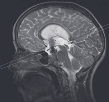
The aim of the study was to determine the possibilities of two differently acquired two-dimensional fast imaging with steady-state precession (FISP 2D) magnetic resonance sequences in estimation of the third ventricle floor fenestration patency after endoscopic third ventriculostomy (ETV) in the subjects with aqueductal stenosis/obstruction.
Fifty eight subjects (37 males, 21 females, mean age 27 years) with previously successfully performed ETV underwent brain MRI on 1.5T MR imager 3-6 months after the procedure. Two different FISP 2D sequences (one included in the standard vendor provided software package, and the other, experimentally developed by our team) were performed respectively at two fixed slice positions: midsagittal and perpendicular to the ETV fenestration, and displayed in a closed-loop cinematographic format in order to estimate the patency. The ventricular volume reduction has been observed as well.
Cerebrospinal fluid (CSF) flow through the ETV fenestration was observed in midsagittal plane with both FISP 2D sequences in 93.11% subjects, while in 6.89% subjects the dynamic CSF flow MRI was inconclusive. In the perpendicular plane CSF flow through the ETV fenestration was visible only by use of experimentally developed FISP 2D (TR30/FA70) sequence. Postoperative volume reduction of lateral and third ventricle was detected in 67.24% subjects.
Though both FISP 2D sequences acquired in midsagittal plane may be used to estimate the effects of performed ETV, due to achieved higher CSF pulsatile flow sensitivity, only the use of FISP 2D (TR30/FA70) sequence enables the estimation of the treatment effect in perpendicular plane in the absence of phase-contrast sequences.
Citations
Downloads
References
Hellwig D, Grotenhuis JA, Tirakotai W, Riegel T, Schulte DM, Bauer BL, et al. Endoscopic third ventriculostomy for obstructive hydrocephalus. Neurosurg Rev. 2005;28(1):1-34
Gangemi M, Maiuri F, Colella G, Magro F, Seneca V, de Divitiis E. Is endoscopic third ventriculostomy an internal shunt alone? Minim Invasive Neurosurg 2007;50(1):47-50
Moorthy RK, Rajshekhar V. Endoscopicthirdventriculostomy for hydrocephalus: a review of indications, outcomes, and complications. Neurol India. 2011;59(6):848-54
Baldauf J, Oertel J, Gaab MR, Schroeder HW. Endoscopic third ventriculostomy in children younger than 2 years of age. Childs Nerv Syst 2007;23:623-6
Farin A, Aryan HE, Ozgur BM, Parsa AT, Levy ML. Endoscopic third ventriculostomy. J Clin Neurosci. 2006 ;13(7):763-70
El-Ghandour NM. Endoscopic third ventriculostomy versus ventriculoperitoneal shunt in the treatment of obstructive hydrocephalus due to posterior fossa tumors in children. Childs Nerv Syst. 2011;27(1):117-26
Hailong F, Guangfu H, Haibin T, Hong P, Yong C, Weidong L, et al. Endoscopic third ventriculostomy in the management of communicating hydrocephalus: a preliminary study. J Neurosurg. 2008;109(5):923-30
Gangemi M, Maiuri F, Buonamassa S, Colella G, de Divitiis E. Endoscopic third ventriculostomy in idiopathic normal pressure hydrocephalus. Neurosurgery. 2004;55(1):129-34
O’Brien DF, Javadpour M, Collins DR, Spennato P, Mallucci CL. Endoscopic third ventriculostomy: an outcome analysis of primary cases and procedures performed after ventriculoperitoneal shunt malfunction. J Neurosurg 2005;103:393-400
Swift D, Nagy L, Robertson B. Endoscopic third ventriculostomy in hydrocephalus associated with achondroplasia. J Neurosurg Pediatr. 2012; 9(1):73-81
Stivaros SM, Sinclair D, Bromiley PA, Kim J, Thorne J, Jackson A. Endoscopic Third Ventriculostomy: Predicting Outcome with Phase-Contrast MR Imaging. Radiology. 2009;252(3):825-32
Hu WD, Xiang L, Wang XR. Cerebrospinal fluid flow in empty sella syndrome and normal sellar regions measured by phase-contrast quantitative magnetic resonance. Neural Regen Res. 2011;6(11):816-20
Dincer A, Yildiz E, Kohan S, Memet Ozek M. Analysis of endoscopic third ventriculostomy patency by MRI: value of different pulse sequences, the sequence parameters, and the imaging planes for investigation of flow void. Childs Nerv Syst. 2011;27(1):127-35
Johanson CE, Duncan JA 3rd, Klinge PM, Brinker T, Stopa EG, Silverberg GD. Multiplicity of cerebrospinal fluid functions: New challenges in health and disease. Cerebrospinal Fluid Res 2008, 5:10
Hoffmann KT, Lehmann TN, Baumann C, Felix R. CSF flow imaging in the management of third ventriculostomy with a reversed fast imaging with steady-state precession sequence. Eur Radiol. 2003;13(6):1432-7
Lucic MA, Koprivsek KM, Till V, Vesic Z. Dynamic magnetic resonance imaging of the cerebrospinal fluid flow within the cerebral aqueduct by different FISP 2D sequences. Vojnosanit Pregl. 2010;67(5): 357-63
Feinberg DA, Mark AS. Human brain motion and cerebrospinal circulation demonstrated with MR velocity imaging. Radiology 1987;163:793-9
Haacke EM, Wielopolski PA, Tkach JA, Modic MT. Steady-state free precession imaging in the presence of motion: application for improved visualization of the cerebrospinal fluid. Radiology 1990; 175:545-52
Quencer RM. Intracranial CSF flow in pediatric hydrocephalus: evaluation with CINE-MR imaging, AJNR Am J Neuroradiol 1992;13:601-8
Santamarta D, Martin-Vallejo J, Díaz-Alvarez A, Maillo A. Changes in ventricular size after endoscopic third ventriculostomy. Acta Neurochir. 2008;150(2):119-127
St George E, Natarajan K, Sgouros S. Changes in ventricular volume in hydrocephalic children following successful endoscopic third ventriculostomy. Childs Nerv Syst. 2004;20(11-12):834-8
Nowosławska E, Polis L, Kaniewska D, Mikołajczyk W, Krawczyk J, Szymański W, et al. Influence of neuroendoscopic third ventriculostomy on the size of ventricles in chronic hydrocephalus. J Child Neurol. 2004;19(8):579-87
Romeo A, Naftel RP, Griessenauer CJ, Reed GT, Martin R, Shannon CN, et al. Long-term change in ventricular size following endoscopic third ventriculostomy for hydrocephalus due to tectal plate gliomas. J Neurosurg Pediatr. 2013;11(1):20-5
Overall WR, Nishimura DG, Hu BS. Fast phase-contrast velocity measurement in the steady state. Magn Reson Med 2002;48(5): 890-8
Scheffler K, Lehnhardt S. Principles and applications of balanced SSFP techniques. Eur Radiol 2003;13(11):2409-2418
Markl M, Pelc NJ. On flow effects in balanced steady-state free precession imaging: pictorial description, parameter dependence, and clinical implications. J Magn Reson Imaging 2004;20(4):697-705
Downloads
Additional Files
Published
Issue
Section
Categories
License
Copyright (c) 2015 Bosnian Journal of Basic Medical Sciences

This work is licensed under a Creative Commons Attribution 4.0 International License.
How to Cite
Accepted 2014-07-24
Published 2014-08-16


