Sectional anatomy of auditory tube
DOI:
https://doi.org/10.17305/bjbms.2004.3406Keywords:
auditory tube, anatomyAbstract
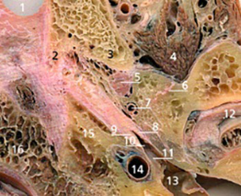
The auditory tube connects the tympanic cavity with nasopharynx. Due to its structure and position, it is difficult to demonstrate the auditory tube in its whole length and to study its topography on anatomical specimens. The purpose of our study was to present the sectional anatomy of the auditory tube in order to facilitate understanding of its structure and topography. We utilized serial sections of the cadaveric head in four planes: transverse, oblique, frontal and sagittal. The osseous part of the auditory tube was demonstrated on transverse sections and most of the cartilaginous part on oblique sections of the head and neck. The tensor veli palati muscle was found to consist of bilaminar muscle sheet: the outer part originating from the skull base and the inner part originating from the lateral cartilaginous lamina and membranous part of the tube. Topographic relations seen on four section planes were described in detail. The structure, course, and topography of auditory tube are well demonstrated on sectional images. Detailed knowledge of the sectional anatomy of the auditory tube is important for the interpretation of corresponding computerized tomographic and magnetic resonance images, and in understanding the disorders and diseases affecting the middle ear and mastoid.
Citations
Downloads
Downloads
Published
How to Cite
Accepted 2018-03-23
Published 2004-05-20


