Direct Immunofluorescence and Immunohistochemistry in Diagnostics of Glomerulonephritis
DOI:
https://doi.org/10.17305/bjbms.2008.2989Keywords:
immunofluorescence (IF), immunoperoxidase (IP), renal biopsies, immune depositsAbstract
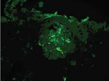
The needle biopsies from 60 transplanted and native kidneys have been processed and a prospective analysis of pattern, intensity and distribution of immunoglobulin deposits (IgA, IgG and IgM) and complement components (C3c and C1q) identified in these lesions has been carried out by immunohistochemistry with three step immunoperoxidase, in the period from 2000 to 2004. Those deposits were previously detected and analyzed by immunofluorescence. The samples consisted of 30 renal biopsies, previously diagnosed with glomerulonephritis and positive immunofluorescence and 30 renal biopsies without morphologic changes and deposits on immunofluorescence. 78,7% of the analyzed samples showed the identical results of the deposits of immunoglobulin and components of the complement with both, immunohistochemistry and immunofluorescence method. Sensitivity of the immunohistochemistry method with three step immunoperoxidase for all analyzed immunoglobulin and complement components is high (0,93), while specificity for the same method is 0,79. Standardized method of the three step immunoperoxidase on the paraffin embedded, formalin fixed needle renal biopsies could successfully replace the immunofluorescence method in diagnostic of GN, with the emphasis on a follow up and control of each single step in the procedure of the method.
Citations
Downloads
Downloads
Published
How to Cite
Accepted 2018-01-15
Published 2008-02-20