The effects of platelet gel on cultured human retinal pigment epithelial (hRPE) cells

Authors

  • Sahar Balagholi Department of Hematology, School of Allied Medicine, Tehran University of Medical Sciences, Tehran, Iran
  • Shaban Alizadeh Department of Hematology, School of Allied Medicine, Tehran University of Medical Sciences, Tehran, Iran
  • Abouzar Bagheri Ocular Tissue Engineering Research Center, Shahid Beheshti University of Medical Sciences, Tehran, Iran; Genetic Research Center, University of Social Welfare and Rehabilitation Sciences, Tehran, Iran
  • Yashar Amizadeh Ocular Tissue Engineering Research Center, Shahid Beheshti University of Medical Sciences, Tehran, Iran
  • Mozhgan Rezaei Kanavi Ocular Tissue Engineering Research Center, Shahid Beheshti University of Medical Sciences, Tehran, Iran http://orcid.org/0000-0002-1497-2260

DOI:

https://doi.org/10.17305/bjbms.2017.2103

Keywords:

Platelet gel, human retinal pigment epithelial cells, PAX6, cell viability, PG, hRPE cells

Abstract

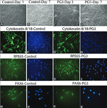
The positive role of platelet gel (PG) in tissue regeneration is well known, however, other characteristics of PG still remain to be determined. We investigated cellular and molecular changes in cultured human retinal pigment epithelial (hRPE) cells when treated with different concentrations of PG named PG1, PG2, and PG3. hRPE cells were isolated from donor eyes of two newborn children, within 24 hours after their death. The cells were treated with three concentrations of PG for 7 days: 3 × 10⁴/ml (PG1), 6 × 10⁴/ml (PG2), and 9 × 10⁴/ml (PG3). Fetal bovine serum was used as a control. Immunocytochemistry was performed with anti-RPE65 (H-85), anti-Cytokeratin 8/18 (NCL-5D3), and anti-PAX6 antibody. We used MTT assay to determine cell viability. Gene expressions of PAX6, MMP2, RPE65, ACTA2, MKI67, MMP9, and KDR were analyzed using real-time PCR. A significant increase in viability was observed for PG3-treated cells compared to control (p = 0.044) and compared to PG1 group (p = 0.027), on day 7. Cellular elongation together with dendritiform extensions were observed in PG-treated cells on days 1 and 3, while epithelioid morphology was observed on day 7. All cells were immunoreactive for RPE65, cytokeratin 8/18, and PAX6. No significant change was observed in the expression of MKI67 and PAX6, but the expressions of MMP2, MMP9, ACTA2, and KDR were significantly higher in PG2-treated cells compared to controls (p < 0.05). Our results indicate that increased concentration of PG and extended exposure time have positive effects on viability of hRPE cells. PG may be useful for hRPE cell encapsulation in retinal cell replacement therapy.

Downloads

Download data is not yet available.

Author Biographies

Sahar Balagholi, Department of Hematology, School of Allied Medicine, Tehran University of Medical Sciences, Tehran, Iran

Department of Hematology

Shaban Alizadeh, Department of Hematology, School of Allied Medicine, Tehran University of Medical Sciences, Tehran, Iran

Department of Hematology

Abouzar Bagheri, Ocular Tissue Engineering Research Center, Shahid Beheshti University of Medical Sciences, Tehran, Iran; Genetic Research Center, University of Social Welfare and Rehabilitation Sciences, Tehran, Iran

Ocular Tissue Engineering Research Center;

Genetic Research Center

Yashar Amizadeh, Ocular Tissue Engineering Research Center, Shahid Beheshti University of Medical Sciences, Tehran, Iran

Ocular Tissue Engineering Research Center

Mozhgan Rezaei Kanavi, Ocular Tissue Engineering Research Center, Shahid Beheshti University of Medical Sciences, Tehran, Iran

Ocular Tissue Engineering Research Center

References

The Eye M.D. Association. Retina and retinal pigment epithelium. In: Ophthalmic Pathology and Intraocular Tumors. Basic and Clinical Science Course. 2016-2017th ed. San Francisco: American Academy of Ophthalmology; 2016. p. 148.

Klein R, Chou CF, Klein BE, Zhang X, Meuer SM, Saaddine JB. Prevalence of age-related macular degeneration in the US population. Arch Ophthalmol 2011;129(1):75-80. https://doi.org/10.1001/archophthalmol.2010.318.

Mehta S. Age-related macular degeneration. Prim Care 2015;42(3):377-91. https://doi.org/10.1016/j.pop.2015.05.009.

Alexander P, Thomson HA, Luff AJ, Lotery AJ. Retinal pigment epithelium transplantation: Concepts, challenges, and future prospects. Eye (Lond) 2015;29(8):992-1002. https://doi.org/10.1038/eye.2015.89.

Forest DL, Johnson LV, Clegg DO. Cellular models and therapies for age-related macular degeneration. Dis Model Mech 2015;8(5):421-7. https://doi.org/10.1242/dmm.017236.

Pasovic L, Eidet JR, Lyberg T, Messelt EB, Aabel P, Utheim TP. Antioxidants improve the viability of stored adult retinal pigment epithelial-19 cultures. Ophthalmol Ther 2014;3(1-2):49-61. https://doi.org/10.1007/s40123-014-0024-9.

Ho W, Tawil B, Dunn JC, Wu BM. The behavior of human mesenchymal stem cells in 3D fibrin clots: Dependence on fibrinogen concentration and clot structure. Tissue Eng 2006;12(6):1587-95. https://doi.org/10.1089/ten.2006.12.1587.

Duong H, Wu B, Tawil B. Modulation of 3D fibrin matrix stiffness by intrinsic fibrinogen-thrombin compositions and by extrinsic cellular activity. Tissue Eng Part A 2009;15(7):1865-76. https://doi.org/10.1089/ten.tea.2008.0319.

van den Dolder J, Mooren R, Vloon AP, Stoelinga PJ, Jansen JA. Platelet-rich plasma: Quantification of growth factor levels and the effect on growth and differentiation of rat bone marrow cells. Tissue Eng 2006;12(11):3067-73. https://doi.org/10.1089/ten.2006.12.3067.

Jalowiec JM, D'Este M, Bara JJ, Denom J, Menzel U, Alini M, et al. An in vitro investigation of platelet-rich plasma-gel as a cell and growth factor delivery vehicle for tissue engineering. Tissue Eng Part C Methods 2016;22(1):49-58. https://doi.org/10.1089/ten.tec.2015.0223.

Najafabadi HS, Soheili ZS, Ganji SM. Behavior of a spontaneously arising human retinal pigment epithelial cell line cultivated on thin alginate film. J Ophthalmic Vis Res 2015;10(3):286-94. https://doi.org/10.4103/2008-322X.170357.

Moroz A, Bittencourt RA, Felisbino SL, da Rosa Pereira H, Rossi-Ferreira R, Deffune E. Platelet gel: 3D scaffold for cell culture. Acta Ortop Bras 2009;17(2):43-5. https://doi.org/10.1590/S1413-78522009000200008.

Heller JP, Martin KR. Enhancing RPE cell-based therapy outcomes for AMD: The role of Bruch's membrane. Transl Vis Sci Technol 2014;3(3):11. https://doi.org/10.1167/tvst.3.4.4.

Falkner-Radler CI, Krebs I, Glittenberg C, Povazay B, Povazay B, Drexler W, et al. Human retinal pigment epithelium (RPE) transplantation: Outcome after autologous RPE choroid sheet and RPE cell-suspension in a randomised clinical study. Br J Ophthalmol 2011;95(3):370-5. https://doi.org/10.1136/bjo.2009.176305.

Binder S. Scaffolds for retinal pigment epithelium (RPE) replacement therapy. Br J Ophthalmol 2011;95(4):441-2. https://doi.org/10.1136/bjo.2009.171926.

Afshari FT, Kwok JC, Andrews MR, Blits B, Martin KR, Faissner A, et al. Integrin activation or alpha 9 expression allows retinal pigmented epithelial cell adhesion on Bruch's membrane in wet age-related macular degeneration. Brain 2010;133(Pt 2):448-64. https://doi.org/10.1093/brain/awp319.

Xie X, Wang Y, Zhao C, Guo S, Liu S, Jia W, et al. Comparative evaluation of MSCs from bone marrow and adipose tissue seeded in PRP-derived scaffold for cartilage regeneration. Biomaterials 2012;33(29):7008-18. https://doi.org/10.1016/j.biomaterials.2012.06.058.

Campochiaro PA, Hackett SF, Vinores SA, Freund J, Csaky C, LaRochelle W, et al. Platelet-derived growth factor is an autocrine growth stimulator in retinal pigmented epithelial cells. J Cell Sci 1994;107(Pt 9):2459-69.

Booij JC, Baas DC, Beisekeeva J, Gorgels TG, Bergen AA. The dynamic nature of Bruch's membrane. Prog Retin Eye Res 2010;29(1):1-18. https://doi.org/10.1016/j.preteyeres.2009.08.003.

Das A, Frank RN, Zhang NL, Turczyn TJ. Ultrastructural localization of extracellular matrix components in human retinal vessels and Bruch's membrane. Arch Ophthalmol 1990;108(3):421-9. https://doi.org/10.1001/archopht.1990.01070050119045.

Ho TC, Del Priore LV. Reattachment of cultured human retinal pigment epithelium to extracellular matrix and human Bruch's membrane. Invest Ophthalmol Vis Sci 1997;38(6):1110-8.

Han J, Yan XL, Han QH, Li YJ, Du ZJ, Hui YN. Integrin ß1 subunit signaling is involved in the directed migration of human retinal pigment epithelial cells following electric field stimulation. Ophthalmic Res 2011;45(1):15-22. https://doi.org/10.1159/000313552.

Tezel TH, Del Priore LV. Reattachment to a substrate prevents apoptosis of human retinal pigment epithelium. Graefes Arch Clin Exp Ophthalmol 1997;235(1):41-7. https://doi.org/10.1007/BF01007836.

Moroz A, Deffune E. Platelet-rich plasma and chronic wounds: Remaining fibronectin may influence matrix remodeling and regeneration success. Cytotherapy 2013;15(11):1436-9. https://doi.org/10.1016/j.jcyt.2013.05.019.

Li H, Usas A, Poddar M, Chen CW, Thompson S, Ahani B, et al. Platelet-rich plasma promotes the proliferation of human muscle derived progenitor cells and maintains their stemness. PLoS One 2013;8(6):e64923. https://doi.org/10.1371/journal.pone.0064923.

Rubio-Azpeitia E, Andia I. Partnership between platelet-rich plasma and mesenchymal stem cells: In vitro experience. Muscles Ligaments Tendons J 2014;4(1):52-62. https://doi.org/10.11138/mltj/2014.4.1.052.

Giusti I, D'Ascenzo S, Mancò A, Di Stefano G, Di Francesco M, Rughetti A, et al. Platelet concentration in platelet-rich plasma affects tenocyte behavior in vitro. Biomed Res Int 2014;2014:630870. https://doi.org/10.1155/2014/630870.

Hotary KB, Yana I, Sabeh F, Li XY, Holmbeck K, Birkedal-Hansen H, et al. Matrix metalloproteinases (MMPs) regulate fibrin-invasive activity via MT1-MMP-dependent and -independent processes. J Exp Med 2002;195(3):295-308. https://doi.org/10.1084/jem.20010815.

Eichler W, Friedrichs U, Thies A, Tratz C, Wiedemann P. Modulation of matrix metalloproteinase and TIMP-1 expression by cytokines in human RPE cells. Invest Ophthalmol Vis Sci 2002;43(8):2767-73.

Moroz A, Delella FK, Lacorte LM, Deffune E, Felisbino SL. Fibronectin induces MMP2 expression in human prostate cancer cells. Biochem Biophys Res Commun 2013;430(4):1319-21. https://doi.org/10.1016/j.bbrc.2012.12.031.

Moroz A, Felisbino SL, Deffune E. Platelet and plasma bioactive scaffolds for stem cell differentiation: What are we missing? Platelets 2014;25(7):556-7. https://doi.org/10.3109/09537104.2013.836748.

Wolf G. Function of the protein RPE65 in the visual cycle. Nutr Rev 2005;63(3):97-100. https://doi.org/10.1111/j.1753-4887.2005.tb00127.x.

Kiser PD, Palczewski K. Membrane-binding and enzymatic properties of RPE65. Prog Retin Eye Res 2010;29(5):428-42. DOI: 10.1016/j.preteyeres.2010.03.002.

Kiser PD, Zhang J, Badiee M, Li Q, Shi W, Sui X, et al. Catalytic mechanism of a retinoid isomerase essential for vertebrate vision. Nat Chem Biol 2015;11(6):409-15. https://doi.org/10.1038/nchembio.1799.

The effects of platelet gel on cultured human retinal pigment epithelial (hRPE) cells

Downloads

Additional Files

Published

2017-11-20

How to Cite

1.
Balagholi S, Alizadeh S, Bagheri A, Amizadeh Y, Rezaei Kanavi M. The effects of platelet gel on cultured human retinal pigment epithelial (hRPE) cells. Biomol Biomed [Internet]. 2017Nov.20 [cited 2023Sep.28];17(4):315-22. Available from: https://bjbms.org/ojs/index.php/bjbms/article/view/2103

Issue

Section

Molecular Biology

INTRODUCTION

The retinal pigment epithelium (RPE) is a layer of tightly connected hexagonal cells, which supports the structural and functional integrity of the photoreceptors and exerts a critical role in the visual process [1]. Age-related macular degeneration (AMD) is a progressive degeneration of the retina and represents the leading cause of blindness worldwide. AMD is characterized by the dysfunction and death of RPE and photoreceptor cells [2,3].

Cultured RPE cells are widely used in AMD research, to investigate the molecular mechanisms of AMD, as well as to test the proposed treatment modalities. Moreover, given the viability of RPE cells in culture media, absence of synaptic connections between the cells and photoreceptors, and easy imaging of the outer retina, novel cell replacement therapies based on these cells have recently emerged for the treatment of AMD [4,5]. In this regard, all attempts are applied to increase the viability and improve the quality of the cells in a culture, to achieve the best results [6].

Platelet gel (PG) obtained from the human blood is a biocompatible and biodegradable natural biopolymer used in tissue engineering. It is the source of several cytokines as well as fibronectin, platelet-derived growth factor (PDGF)-AA, -BB, and -AB, transforming growth factor b (TGF-b) 1 and 2, platelet-derived epidermal growth factor (PDEGF), platelet-derived angiogenesis factor (PDAF), insulin-like growth factor-1 (IGF-1), and platelet factor-4 (PF-4). Due to its rich content, PG is proposed to be used as a three-dimensional (3D) structure for the cultivation of different cells [7-10]. However, apart from the positive role of PG in cell cultivation, other characteristics still remain to be determined. In this study, we evaluated the effect of three different concentrations of PG on human RPE (hRPE) cells, in vitro. Our findings could provide valuable information in the treatment planning of ocular disorders that are difficult to treat and in need for transplantation of healthy viable cells.

MATERIALS AND METHODS

Donor eyes of two newborn children (6 and 8 months old) were obtained from the Central Eye Bank of Iran, within 24 hours after their death. The research approval was obtained from the Institutional Review Board of the Central Eye Bank of Iran and the Ethics Committee of the Ophthalmic Research Center at the Shahid Beheshti University of Medical Sciences, Tehran, Iran.

Isolation and cultivation of hRPE cells

The isolation and cultivation of hRPE cells were performed as described previously [11]. Briefly, the posterior parts of the donor eyes were obtained from the Central Eye Bank of Iran, Tehran, Iran. After the excision of the corneosclera for transplantation purposes, the remaining posterior part of the eye was stored at 4°C in a moist chamber containing antibiotics, shipped immediately to the cell culture laboratory, and dissected on the same day of shipping. The dissection of the posterior eye cup was initiated by cutting off the iris/lens complex carefully and removing the vitreous body. After dissecting the posterior eye cup into four quadrants, the sensory retina was removed and the RPE/choroid layer was gently peeled of the sclera and incubated in 2% dispase solution for an hour at 37°C, with 5% CO2. Then, the tissue was irrigated with phosphate-buffered saline (PBS) and the isolated RPE sheets were cultured in T75 flasks (Nunc, Roskilde, Denmark) containing Dulbecco’s modified Eagle’s medium/Ham’s F12 [DMEM/F12] (Sigma-Aldrich, Munich, Germany) supplemented with 20% fetal bovine serum [FBS] (GIBCO-BRL, Eggenstein, Germany) and incubated at 37°C, with 5% CO2. The experiments were performed using hRPE cells that were passaged 3-5 times.

Preparation of PG and treatment of cultured hRPE cells

A citrated blood sample with 3 × 10⁵/dL platelets was provided from a healthy volunteer and centrifuged at 130 g for 15 minutes, at 25°C. Next, thrombin was applied to activate the platelets. The platelet rich plasma (PRP), with three different concentrations of platelets (3 × 10⁴/ml [PG1], 6 × 10⁴/ml [PG2], and 9 × 10⁴/ml [PG3]), was added into each well of a 96-well cell culture plate. Thereafter, thrombin was added at a rate of 5% of the final volume. The resultant product was a gel that remained solid at both room (25°C) and body (37°C) temperatures. When the gel was formed, 1000 hRPE cells were seeded into each well. The wells that were not treated with PG and containing 20% FBS were considered as controls.

Immunocytochemical characterization of PG-treated hRPE cells

The hRPE cells passaged 3-5 times and with the density of 5 × 10³ cells/well were encapsulated with the three different concentrations of PG, in 24-well microplates (Nunc, Roskilde, Denmark), and incubated at 37°C, with 5% CO2, for 24 hours. The non-treated cells were considered as controls. After 7 days, following the irrigation of the wells with PBS, the hRPE cells were fixed with ice-cold methyl alcohol [−10°C] (Merck, Darmstadt, Germany) for 5 minutes at room temperature. Then, the slides were blocked with a blocking solution containing 1% bovine serum albumin [BSA] (Merck, Darmstadt, Germany) in 1% Triton X-100 in PBS (Sigma-Aldrich, Munich, Germany) for 20 minutes at room temperature. After removing the blocking agent and irrigating with PBS, the wells were incubated with diluted solution of anti-RPE65 antibody [H-85] (rabbit polyclonal immunoglobulin G [IgG], Santa Cruz Biotechnology Inc., Dallas, USA), anti-cytokeratin 8/18 antibody [NCL-5D3] (mouse monoclonal IgG2a, Santa Cruz Biotechnology Inc., Dallas, USA), and anti-PAX6 antibody (goat polyclonal IgG, Santa Cruz Biotechnology Inc., Dallas, USA) overnight at 4°C. Following irrigation with PBS, fluorescein isothiocyanate (FITC)-conjugated goat anti-rabbit IgG (1:100, Santa Cruz, Carlsbad, CA, USA), FITC-conjugated goat anti-mouse IgG (1:100, Santa Cruz, Carlsbad, CA, USA), and FITC-conjugated donkey anti-goat IgG (1:100, Santa Cruz, Carlsbad, CA, USA) were applied, respectively, on the coverslips incubated with H-85, NCL-5D3, and anti-PAX6, for 45 minutes in the dark and at room temperature. Subsequently, the cells were stained with 4’, 6-diamidino-2-phenylindole [DAPI] (1 mg/ml, Santa Cruz, Carlsbad, CA, USA) for 5 minutes and examined under an inverted microscope (Axiophot, Zeiss, Germany), equipped with a 460 nm filter for DAPI and a 520 nm filter for FITC-conjugated antibodies.

Cell viability assay

To test the cell viability, 3-(4,5-dimethylthiazol-2-yl)-2,5-diphenyltetrazolium bromide [MTT] (Sigma-Aldrich, Munich, Germany) was added to the cell culture wells at a rate of 10% of the initial volume and the solution was incubated for 4 hours. Then, the medium and MTT were removed and 100 ml of dimethyl sulfoxide (DMSO) was added into each well. The absorbance was read by an enzyme-linked immunosorbent assay (ELISA) analyzer (BioTek Instruments, Inc, USA) at a wavelength of 540 nm.

RNA isolation

The total RNA was extracted from hRPE cells with TRIzol reagent (Ambion, USA). The samples were then reacted at room temperature for 5 minutes. Chloroform was added to further separate RNA from other cell components and 500 ml of isopropanol was added to precipitate RNA. The RNA was then washed with 75% EtOH and dissolved in nuclease-free water. The concentration and purity of the isolated RNA were determined by a NanoDrop spectrophotometer (Thermo Fisher Scientific, USA) and the integrity of the RNA was assessed with agarose gel electrophoresis.

Quantitative reverse transcription polymerase chain reaction (qRT-PCR)

The RT reaction was performed with oligo (dT) primers and a SuperScript reverse transcriptase kit (Promega, USA). Then, qPCR was performed using an EvaGreen qPCR master mix (Solis BioDyne, Estonia). The PCR parameters were as follows: initial denaturation for one cycle at 95°C, for 15 minutes; denaturation, amplification, and quantification for 40 cycles at 95°C for 15 seconds, 56-64°C for 17 seconds, and 72°C for 25 seconds, respectively; generation of the melting curve at 65°C, with the temperature gradually increasing to 95°C. The mRNA expression was normalized to the levels of glyceraldehyde 3-phosphate dehydrogenase (GAPDH) mRNA, and the efficiency (E) for each primer was calculated according to the slope of the standard curve. The primer sequences used for qPCR are provided in Table 1.

TABLE 1: Primer sequences

Statistical analysis

All statistical analyses were performed using IBM SPSS Statistics for Windows, Version 22.0 (IBM Corp., Armonk, NY). The qRT-PCR analysis was performed at least in three independent experiments. Each sample was run and analyzed in duplicate. Differences between the groups were analyzed using non-parametric Kruskal–Wallis test and multiple comparison tests. A value of P < 0.05 was considered statistically significant.

RESULTS

Morphological and immunocytochemical analysis of PG-treated hRPE cells

Cellular elongation together with dendritiform extensions were observed in PG-treated cells on days 1 and 3 [Figure 1]. However, on day 7, following the complete digestion of PG, the cells migrated toward the two-dimensional surface of the wells and revealed predominantly epithelioid morphology. The digestion of platelet gel led to the formation of insoluble residues in the culture medium. Since our experiments were performed with cultured cells passaged 3-5 times, marked pigmentation was not observed in the treated or control cells. All PG-treated cells were immunoreactive for cytokeratin 8/18 (Figure 1E), RPE65 (Figure 1I), and PAX6 (Figure 1M), and this was not significantly different from the corresponding controls (Figure 1G, K, and O, respectively).

FIGURE 1: Photomicrographs of cultured human retinal pigment epithelial (hRPE) cells seeded on platelet gel of 9 × 10⁴/ml concentration (PG3) versus controls. Note the epithelioid morphology of the control cells on days 3 (A) and 7 (B), and elongated morphology with dendriform extensions of PG3-treated cells on day 3 (C). The PG3-treated cells demonstrated epithelioid morphology on day 7 (D). Both control (E, I, M) and PG3-treated cells (G, K, O) were positively stained (green) for the fluorescein isothiocyanate (FITC)-conjugated cytokeratin 8/18 antibody (E and G), FITC-conjugated retinal pigment epithelium-specific protein 65 kDa (RPE65) antibody (I and K), and FITC-conjugated paired box protein-6 (PAX6) antibody (M and O). Note 4’, 6-diamidino-2-phenylindole (DAPI)-stained, hRPE cell nuclei (blue) in images F, J, and N for the controls, and in images H, L, and P for PG3-treated cells. The magnification is × 200 in all images.

Viability of PG-treated hRPE cells

On days 1 and 3, increased viability of PG-treated hRPE cells was observed compared to the controls, however, this increase was not statistically significant. On day 7, PG3-treated cells showed a significant increase in viability compared to control (P = 0.044) and PG1 group (P = 0.027) [Figure 2].

FIGURE 2: Viability of cultured human retinal pigment epithelial (hRPE) cells 24 hours, 72 hours, and 1 week after the treatment with different concentrations of platelet gel (3 × 10⁴/ml [PG1], 6 × 10⁴/ml [PG2], and 9 × 10⁴/ml [PG3]). Note the increased viability of hRPE cells treated with PG compared to the controls, on days 1 and 3. A higher viability of hRPE cells is observed after PG3 treatment on day 7, as compared to control and PG1-treated cells (*p < 0.05).

Gene expression of PG-treated hRPE cells

According to the qRT-PCR results [Figure 3], neither of the three concentrations of PG had a significant effect on the gene expressions of MKI67 (encodes antigen KI-67 important for cellular proliferation), PAX6 (encodes the transcription factor paired box protein Pax-6), and RPE65 (encodes retinal pigment epithelium-specific 65 kDa protein, an RPE-specific protein). On the other hand, the expressions of MMP2, MMP9 (members of metalloproteinase gene family that encode enzymes involved in the degradation of extracellular matrix proteins), ACTA2 (encodes smooth muscle alpha [a]-2 actin, a marker of myofibroblast differentiation), and KDR (encodes vascular endothelial growth factor receptor 2 [VEGFR-2]) were significantly higher in PG2-treated cells compared to controls (P < 0.05).

FIGURE 3: Gene expression analysis of cultured human retinal pigment epithelial (hRPE) cells exposed to different concentrations of platelet gel (3 × 10⁴/ml [PG1], 6 × 10⁴/ml [PG2], and 9 × 10⁴/ml [PG3]). The illustrated graphs depict the gene expressions of RPE65, PAX6, MKI67, KDR, MMP2, MMP9, and ACTA2 in PG-treated hRPE cells compared to the controls. Note the high expressions of MMP2, MMP9, ACTA2, and KDR genes in PG2-treated cells (*p < 0.05). RPE65: Retinal pigment epithelium-specific protein 65 kDa; PAX6: Paired box protein-6; MKI67: Marker of proliferation Ki-67; MMP-2: Matrix metalloproteinase-2; MMP-9: matrix metalloproteinase-9; ACTA2: Alpha-actin-2; KDR: Kinase insert domain receptor.

DISCUSSION

Our study demonstrated for the first time that increased concentrations of PG and extended exposure time have positive effects on the viability of hRPE cells. Moreover, the gene expression analysis showed that PG did not induced neither tumorigenesis nor reprogramming of the hRPE cells. However, the expression of angiogenic-, myofibroblast-, and inflammation-related genes in PG2-treated hRPE cells was significantly higher compared to the controls. Nevertheless, these transcriptional changes were not significant in PG1- and PG3-treated cells.

Platelets obtained from the human blood are a good source of several cytokines and growth factors, and PG can be used as a 3D scaffold for cell culture [12]. Consistent with this information, in our study, the viability of PG-treated hRPE cells increased over the 1-week period, especially in PG3-treated cells on day 7, compared to control group. These results indicate that a 1-week exposure of hRPE cells to 9 × 10⁴/ml of PG have a considerable impact on the growth and function of hRPE cells. In addition, this effect of PG on cultured hRPE cells might be due to the partial encapsulation of the cells by the gel, thus functioning as an appropriate scaffold for cell survival.

PG has essential, structural and compositional, similarities to the extracellular matrix (ECM). In addition to providing essential nutrients, it enables free exchange of nutrients and oxygen between the encapsulated cells and their surrounding environment. This characteristic of PG makes it an ideal candidate as a vehicle for hRPE cell delivery and a temporary support for RPE regeneration during transplantation in AMD, to replace the damaged RPE cells [2]. However, it remains to be investigated whether an autologous PG can reduce the immunogenicity of non-autologous hRPE cells.

In RPE cell replacement therapy for dry AMD, the adherence of the cells to the extracellular environment plays a pivotal role in the survival of grafted cells [13]. Although no significant difference was observed between RPE cell-suspension and RPE-choroid sheet transplantation techniques, the apoptosis of RPE cells is more likely to occur after the transplantation of the cell suspensions into the pathological environment [14,15]. Therefore, different types of tissue-engineered scaffolds have been used to increase the binding of transplanted cells to the damaged tissue, while enabling the cells to cope with the surrounding matrix [16]. One of the challenges with these substrates is the occurrence of post-transplantation wrinkling or contraction of synthetic scaffolds that were used for RPE grafts [15]. As an autologous, highly nutrient and biodegradable scaffold, we propose PG as a potentially ideal substrate for easy injection and transplantation of RPE cell suspensions. Moreover, the presence of bioactive factors in PG, such as PDGF and TGF-b, which are autocrine stimulators of RPE cell growth [17,18], may not only be effective in healing the damaged tissue but may also enhance the attachment of the grafted RPE cells and improve their functionality. Further studies should investigate the accuracy of our assumptions.

Pathological changes in Bruch’s membrane, a distinctive feature of AMD, is one of the challenges encountered in RPE cell transplantation. Extracellular ligands, such as laminin and fibronectin, that are present in the uppermost layers of Bruch’s membrane, mediate the binding of RPE cells to Bruch’s membrane and also inhibit the cell apoptosis [16,19-23]. As a rich source of these extracellular ligands [24], PG may significantly enhance the proper attachment of grafted RPE cells.

In the current study, the hRPE cells exposed to PG did not show multipotent capacity at the level of gene expression. Our results are consistent with the study of Li et al. [25], in which PG significantly enhanced the short- and long-term proliferation of human muscle-derived stem cells, without altering the differentiation capacity of the cells. The same findings were obtained by Rubio-Azpeitia et al. [26], in which PG stimulated human mesenchymal stem cell (hMSC) proliferation and preserved their multipotency; moreover, it did not interfere with any lineage differentiation. Also, the PG maintained the immunomodulatory properties of hMSCs [26].

In a series of studies, autologous PG reduced the expression of matrix metalloproteinase-2 (MMP-2), while it promoted the expression of tissue inhibitor of MMP-2 (TIMP-2) [6]. Giusti et al. [27] showed that the highest concentration of platelet-rich plasma [PRP] (5 × 10⁶ plt/mL) induced the MMP production and proteolysis in cultured tenocytes. In our study, the gene expression analysis of cultured hRPE cells, exposed to different concentrations of PG, showed the upregulation of MMP2 and MMP9 genes in PG2- as compared with PG1- and PG3-treated cells. In other studies, the digestion of PG has been demonstrated as the result of MMP secretion, from cultured RPE cells [28,29]. The digestion of PG, in our study, may also be related to the increased expression of MMP2 and MMP9 genes in the PG-treated hRPE cells. Furthermore, the observed high expression of the MMP genes might be a physiological response of the PG-treated hRPE cells, and could be beneficial for PG absorption and remodeling. The digestion of PG in our cultures led to the formation of insoluble, but biocompatible, residues in the culture medium, which might have originated from the degradation of PG fibrin fibers. It has been suggested that the MMP activity increases after short-time exposure to fibronectin that is present in PRP-based scaffolds, and this could be beneficial for transdifferentiation process in cell replacement therapies [30,31].

Neither of the three concentrations of PG, in our study, had a significant effect on the expression of MKI67 and PAX6 genes. Moreover, the expression of PAX6 protein was not significantly different from the controls, based on the immunocytochemical analysis. On the other hand, PG2-treated hRPE cells showed a significant increase in the expression of ACTA2 gene. The increased viability of PG3-treated hRPE cells, with no significant changes in the expression of MKI67 and PAX6 genes, and with comparable expression of PAX6 protein to the controls, make PG a promising 3D substrate, with no risk of tumorigenicity, myofibroblastic differentiation, and cell reprogramming, for the treated hRPE cells. The related in vivo studies should be conducted to investigate the potential of PG in ocular, cell-based therapies.

The KDR gene, encoding VEGFR-2, was significantly amplified in our PG2-treated hRPE cells, which is not favorable for angiogenesis. The expression of KDR was the lowest in PG3-treated hRPE cells compared to PG1- and PG2-treated cells, therefore, the PG concentration of 9 × 10⁴/ml might be appropriate to avoid altered vasculature in the subretinal space, after RPE transplantation.

The RPE65 gene encodes retinal pigment epithelium-specific 65 kDa protein, also known as retinoid isomerohydrolase, which is located in RPE cells. During phototransduction, RPE65 protein is responsible for the conversion of all-trans-retinyl esters to 11-cis-retinol. 11-cis-retinol is then used in visual pigment regeneration in photoreceptor cells [32-34]. In the current study, the expression of RPE65 gene did not significantly change in PG-treated hRPE cells. The qRT-PCR results were also supported by the immunocytochemistry. Whether different concentrations of PG affect the functionality of hRPE cells during phototransduction, needs further comprehensive investigations at the protein level as well as in vivo studies.

CONCLUSION

In summary, our results indicate that PG as a 3D substrate and rich source of cytokines, nutrients and growth factors has a positive effect on the viability of cultured hRPE cells. Furthermore, given the effects of the triple concentration of PG on the gene expression of cultured hRPE cells, the PG concentration of 9 × 10⁴/ml may be suitable for the encapsulation and delivery of hRPE cells in regenerative medicine for retinal diseases.

Acknowledgements

ACKNOWLEDGMENTS

The authors would like to thank the Central Eye Bank of Iran for providing the donor eyes.

DECLARATION OF INTERESTS

The authors declare no conflict of interests.

REFERENCES

  1. (). . Retina and retinal pigment epithelium. In: Ophthalmic Pathology and Intraocular Tumors. Basic and Clinical Science Course.
  2. , , , , , (). Prevalence of age related macular degeneration in the US population. Arch Ophthalmol. https://doi.org/10.1001/archophthalmol. 2010.318
  3. (). Age related macular degeneration. Prim Care. https://doi.org/10.1016/j.pop. 2015.05.009
  4. , , , (). Retinal pigment epithelium transplantation: concepts, challenges, and future prospects. Eye (Lond). https://doi.org/10.1038/eye. 2015.89
  5. , , (). Cellular models and therapies for age related macular degeneration. Dis Model Mech. https://doi.org/10.1242/dmm. 017236
  6. , , , , , (). Antioxidants improve the viability of stored adult retinal pigment epithelial 19 cultures. Ophthalmol Ther. https://doi.org/10.1007/s40123 014 0024 9
  7. , , , (). The behavior of human mesenchymal stem cells in 3D fibrin clots: dependence on fibrinogen concentration and clot structure. Tissue Eng. https://doi.org/10.1089/ten. 2006.12.1587
  8. , , (). Modulation of 3D fibrin matrix stiffness by intrinsic fibrinogen thrombin compositions and by extrinsic cellular activity. Tissue Eng Part A. https://doi.org/10.1089/ten.tea. 2008.0319
  9. , , , , (). Platelet rich plasma: quantification of growth factor levels and the effect on growth and differentiation of rat bone marrow cells. Tissue Eng. https://doi.org/10.1089/ten. 2006.12.3067
  10. , , , , , (). Anin vitroinvestigation of platelet rich plasma gel as a cell and growth factor delivery vehicle for tissue engineering. Tissue Eng Part C Methods. https://doi.org/10.1089/ten.tec. 2015.0223
  11. , , (). Behavior of a spontaneously arising human retinal pigment epithelial cell line cultivated on thin alginate film. J Ophthalmic Vis Res. https://doi.org/10.4103/2008 322X.170357
  12. , , , , , (). Platelet gel: 3D scaffold for cell culture. Acta Ortop Bras. https://doi.org/10.1590/S1413 78522009000200008
  13. , (). Enhancing RPE cell based therapy outcomes for AMD: The role of Bruch’s membrane. Transl Vis Sci Technol. https://doi.org/10.1167/tvst. 3.4.4
  14. , , , , , (). Human retinal pigment epithelium (RPE) transplantation: Outcome after autologous RPE choroid sheet and RPE cell suspension in a randomised clinical study. Br J Ophthalmol. https://doi.org/10.1136/bjo. 2009.176305
  15. (). Scaffolds for retinal pigment epithelium (RPE) replacement therapy. Br J Ophthalmol. https://doi.org/10.1136/bjo. 2009.171926
  16. , , , , , (). Integrin activation or alpha 9 expression allows retinal pigmented epithelial cell adhesion on Bruch’s membrane in wet age related macular degeneration. Brain. https://doi.org/10.1093/brain/awp319
  17. , , , , , (). Comparative evaluation of MSCs from bone marrow and adipose tissue seeded in PRP derived scaffold for cartilage regeneration. Biomaterials. https://doi.org/10.1016/j.biomaterials. 2012.06.058
  18. , , , , , (). Platelet derived growth factor is an autocrine growth stimulator in retinal pigmented epithelial cells. J Cell Sci.
  19. , , , , (). The dynamic nature of Bruch’s membrane. Prog Retin Eye Res. https://doi.org/10.1016/j.preteyeres. 2009.08.003
  20. , , , (). Ultrastructural localization of extracellular matrix components in human retinal vessels and Bruch’s membrane. Arch Ophthalmol. https://doi.org/10.1001/archopht. 1990.01070050119045
  21. , (). Reattachment of cultured human retinal pigment epithelium to extracellular matrix and human Bruch’s membrane. Invest Ophthalmol Vis Sci.
  22. , , , , , (). Integrin ß1 subunit signaling is involved in the directed migration of human retinal pigment epithelial cells following electric field stimulation. Ophthalmic Res. https://doi.org/10.1159/000313552
  23. , (). Reattachment to a substrate prevents apoptosis of human retinal pigment epithelium. Graefes Arch Clin Exp Ophthalmol. https://doi.org/10.1007/BF01007836
  24. , (). Platelet rich plasma and chronic wounds: remaining fibronectin may influence matrix remodeling and regeneration success. Cytotherapy. https://doi.org/10.1016/j.jcyt. 2013.05.019
  25. , , , , , (). Platelet rich plasma promotes the proliferation of human muscle derived progenitor cells and maintains their stemness. PLoS One. https://doi.org/10.1371/journal.pone.0064923
  26. , (). Partnership between platelet rich plasma and mesenchymal stem cellsin vitroexperience. Muscles Ligaments Tendons J. https://doi.org/10.11138/mltj/2014.4.1.052
  27. , , , , , (). Platelet concentration in platelet rich plasma affects tenocyte behaviorin vitro. Biomed Res Int. https://doi.org/10.1155/2014/630870
  28. , , , , , (). Matrix metalloproteinases (MMPs) regulate fibrin invasive activity via MT1 MMP dependent and independent processes. J Exp Med. https://doi.org/10.1084/jem. 20010815
  29. , , , , (). Modulation of matrix metalloproteinase and TIMP 1 expression by cytokines in human RPE cells. Invest Ophthalmol Vis Sci.
  30. , , , , (). Fibronectin induces MMP2 expression in human prostate cancer cells. Biochem Biophys Res Commun. https://doi.org/10.1016/j.bbrc. 2012.12.031
  31. , , (). Platelet and plasma bioactive scaffolds for stem cell differentiation: what are we missing?. Platelets. https://doi.org/10.3109/09537104.2013.836748
  32. (). Function of the protein RPE65 in the visual cycle. Nutr Rev. https://doi.org/10.1111/j.1753.4887.2005.tb00127.x
  33. , (). Membrane binding and enzymatic properties of RPE65. Prog Retin Eye Res. DOI: 10.1016/j.preteyeres.2010.03.002
  34. , , , , , (). Catalytic mechanism of a retinoid isomerase essential for vertebrate vision. Nat Chem Biol. https://doi.org/10.1038/nchembio. 1799