Significance of Foxp3+CD4+ regulatory T cells in the peripheral blood of Uygur patients in the acute and chronic phases of pigeon breeder’s lung

Authors

  • Biqing Yu Xinjiang Medical University, Urumqi, China; Department of Respiratory and Critical Care Medicine, People’s Hospital of Xinjiang Uygur Autonomous Region, Urumqi, China
  • Xiaohong Yang Department of Respiratory and Critical Care Medicine, People’s Hospital of Xinjiang Uygur Autonomous Region, Urumqi, China
  • Fengsen Li Respiratory Department, Hospital of Traditional Chinese Medicine of Xinjiang Uygur Autonomous Region, Urumqi, China
  • Chao Wu Department of Respiratory and Critical Care Medicine, People’s Hospital of Xinjiang Uygur Autonomous Region, Urumqi, China
  • Wenyi Wang Department of Respiratory and Critical Care Medicine, People’s Hospital of Xinjiang Uygur Autonomous Region, Urumqi, China
  • Wei Ding Department of Respiratory and Critical Care Medicine, People’s Hospital of Xinjiang Uygur Autonomous Region, Urumqi, China

DOI:

https://doi.org/10.17305/bjbms.2016.1233

Keywords:

Pigeon breeder’s lung, Foxp3 CD4 Treg, T lymphocyte, Uygur

Abstract

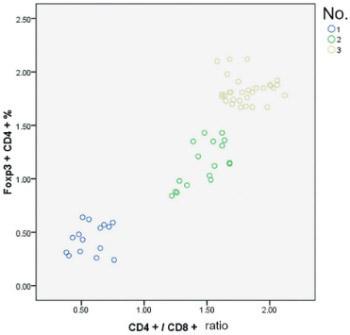
Pigeon breeder’s lung (PBL) is a type of lung inflammatory disease associated with the immune response to repeated pigeon-derived antigen exposure. The pathogenesis of PBL remains unclear. In this study, peripheral blood samples were collected from Uygur acute - and chronic-phase PBL patients and healthy subjects with pigeon contact. Foxp3+CD4+ regulatory T cell (Treg) activity in different phases of PBL was characterized by changes in Foxp3+CD4+ Treg, CD4+CD25+ T cell, and T lymphocyte subsets. Based on hypersensitivity pneumonitis (HP) diagnosis criteria, 32 PBL cases from January 2012 to December 2013 in the People’s Hospital of Xinjiang Uygur Autonomous Region Respiratory Department were included. Lung high-resolution computed tomography was performed, and the cases were classified based on the HP phase into 15 acute-phase and 17 chronic-phase cases. The control group included 30 healthy subjects with Uygur pigeon contact. Blood samples were collected, and the T cell subsets were analyzed via flow cytometry. In both PBL groups, the Foxp3+CD4+ Treg and CD4+CD25+ and CD4+CD3+ T cell percentages and CD4+/CD8+ ratios were significantly lower than in the control group (p < 0.01). In the PBL groups, particularly the acute-phase group, the CD8+CD3+ T lymphocyte percentage was significantly higher than in the control group (p < 0.01). There were no significant differences in CD4+CD25+ cells between the PBL groups. In peripheral blood from the PBL groups, the CD4+/CD8+ ratio was positively correlated with the Foxp3+CD4+ Treg (r = 0.864, p < 0.05) and CD4+/CD25+ cell (r = 0.34, p < 0.05) percentages. Low Foxp3+CD4+ Treg expression or overconsumption may be a pathogenic factor in PBL.

Downloads

Download data is not yet available.

Author Biographies

Biqing Yu, Xinjiang Medical University, Urumqi, China; Department of Respiratory and Critical Care Medicine, People’s Hospital of Xinjiang Uygur Autonomous Region, Urumqi, China

Department of Respiratory and Critical Care Medicine

Xiaohong Yang, Department of Respiratory and Critical Care Medicine, People’s Hospital of Xinjiang Uygur Autonomous Region, Urumqi, China

Department of Respiratory and Critical Care Medicine

Fengsen Li, Respiratory Department, Hospital of Traditional Chinese Medicine of Xinjiang Uygur Autonomous Region, Urumqi, China

Respiratory Department

Chao Wu, Department of Respiratory and Critical Care Medicine, People’s Hospital of Xinjiang Uygur Autonomous Region, Urumqi, China

Department of Respiratory and Critical Care Medicine

Wenyi Wang, Department of Respiratory and Critical Care Medicine, People’s Hospital of Xinjiang Uygur Autonomous Region, Urumqi, China

Department of Respiratory and Critical Care Medicine

Wei Ding, Department of Respiratory and Critical Care Medicine, People’s Hospital of Xinjiang Uygur Autonomous Region, Urumqi, China

Department of Respiratory and Critical Care Medicine

References

. Barrera L, Mendoza F, Zuñiga J, Estrada A, Zamora AC, Melendro EI, et al. Functional diversity of T-cell subpopulations in subacute and chronic hypersensitivity pneumonitis. Am J Respir Crit Care Med 2008;177(1):44-55.

http://dx.doi.org/10.1164/rccm.200701-093OC.

. Dalphin JC, Didier A. Environmental causes of the distal airways disease. Hypersensitivity pneumonitis and rare causes. Rev Mal Respir 2013;30(8):669-81. http://dx.doi.org/10.1016/j.rmr.2013.03.001.

. Camarena A, Aquino-Galvez A, Falfán-Valencia R, Sánchez G, Montaño M, Ramos C, et al. PSMB8 (LMP7) but not PSMB9 (LMP2) gene polymorphisms are associated to pigeon breeder's hypersensitivity pneumonitis. Respir Med 2010;104(6):889-94. http://dx.doi.org/10.1016/j.rmed.2010.01.014.

. Bourke SJ, Dalphin JC, Boyd G, McSharry C, Baldwin CI, Calvert JE. Hypersensitivity pneumonitis: Current concepts. Eur Respir J Suppl 2001;32:81s-92s.

. Girard M, Israël-Assayag E, Cormier Y. Pathogenesis of hypersensitivity pneumonitis. Curr Opin Allergy Clin Immunol 2004;4(2):93-8.

http://dx.doi.org/10.1097/00130832-200404000-00004.

. Bracken SJ, Adami AJ, Szczepanek SM, Ehsan M, Natarajan P, Guernsey LA, et al. Long-term exposure to house dust mite leads to the suppression of allergic airway disease despite persistent lung inflammation. Int Arch Allergy Immunol 2015;166(4):243-58. http://dx.doi.org/10.1159/000381058.

. Qiu SL, Bai J, Zhong XN. CD4+ Foxp3+ regulatory T cells in inflammation and emphysema after smoking cessation in rats. [Article in Chinese]. Chinese Journal of Tuberculosis and Respiratory Diseases. 2010;33(9):688-92.

. Wang LL, Ling Y, Liu S. The effect of Foxp3~+CD4~+ Treg cells in the acute and chronic greenhouse farmer’s lung. China J Mod Med 2012;22(28):40-5.

. Wang W, Wei D, Yang X. Interleukin 10 gene single nucleotide polymorphism and lung Uygur who feed pigeons. Int J Respir 2015;35(18):1401-7.

. Wu C, Chen Y, Yang X, Wang W, Pang B. Correlation of macrophage inflammatory protein-1alpha single gene polymorphisms with the susceptibility to pigeon breeder's lung in Chinese Uygur population. Int J Clin Exp Med 2015;8(8):13732-9.

. Chen JH, Juan H. Profiles of cytokines and their gene polymorphisms in the pathogenesis of pigeon breeder's lung. Curr Immunol 2004;24(3):198-202.

. Muto S, Owada Y, Inoue T, Watanabe Y, Yamaura T, Fukuhara M, et al. Clinical significance of expanded Foxp3+ Helios- regulatory T cells in patients with non-small cell lung cancer. Int J Oncol 2015;47(6):2082-90. http://dx.doi.org/10.3892/ijo.2015.3196.

. Krogulska A, Polakowska E, Wasowska-Królikowska K, Malachowska B, Mlynarski W, Borowiec M. Decreased FOXP3 mRNA expression in children with atopic asthma and IgE-mediated food allergy. Ann Allergy Asthma Immunol 2015;115(5):415-21. http://dx.doi.org/10.1016/j.anai.2015.08.015.

. Schuyler M, Cormier Y. The diagnosis of hypersensitivity pneumonitis. Chest 1997;111(3):534-6. http://dx.doi.org/10.1378/chest.111.3.534.

. Lou CY, Li M, Li L. Dynamic changes in percentages of CD4+ CD25+ regulatory T cells and Th17 cells in process of airway remodeling in mouse model of asthma. [Article in Chinese]. Chin J Contemp Pediatric 2015;17(9):994-1000.

. Maggi L, Santarlasci V, Liotta F, Frosali F, Angeli R, Cosmi L, et al. Demonstration of circulating allergen-specific CD4+CD25(high)FoxP3+ T-regulatory cells in both nonatopic and atopic individuals. J Allergy Clin Immunol 2007;120(2):429-36. http://dx.doi.org/10.1016/j.jaci.2007.05.002.

. Li S, Li Y, Qu X, Liu X, Liang J. Detection and significance of TregFoxP3+ and Th17 cells in peripheral blood of non-small cell lung cancer patients. Arch Med Sci 2014;10(2):232-9. http://dx.doi.org/10.5114/aoms.2014.42573.

. Conte E, Gili E, Fruciano M, Fagone E, Vancheri C. Human lung fibroblasts increase CD4+CD25+Foxp3+ T cells in co-cultured CD4+ lymphocytes. Cell Immunol 2013;285(1-2):55-61. http://dx.doi.org/10.1016/j.cellimm.2013.09.002.

. Chaput N, Marabelle A. Foxp3 cells are running the show in patients with surgically resected nonsmall cell lung cancer. Eur Respir J 2015;46(6):1541-3. http://dx.doi.org/10.1183/13993003.01595-2015.

. Wang LL, Zhao MJ, Mao ST. The relevance of changes of the CD4+ Foxp3+ regular T cells from different stages of elder COPD patients and the lung function. [Article in Chinese]. Chin J Gerontol 2012;32(2):223-5.

. Shi Y, Chu LL, Zhang LF. Role of CD4+CD25+ regulation cell s and expressing of FOXP3 in the pathogenesis of children with asthma. [Article in Chinese]. Acta Univ Med Nanjing 2009;29(1):117-9.

. Lee MG, Bae SC, Lee YH. Association between FOXP3 polymorphisms and susceptibility to autoimmune diseases: A meta-analysis. Autoimmunity 2015;48(7):445-52. http://dx.doi.org/10.3109/08916934.2015.1045582.

. Koschel DS, Cardoso C, Wiedemann B, Höffken G, Halank M. Pulmonary hypertension in chronic hypersensitivity pneumonitis. Lung 2012;190(3):295-302. http://dx.doi.org/10.1007/s00408-011-9361-9.

. Shi HY, Deng WJ, Zhang YP, Zhong YJ, Liu J, Fang P, et al. Expression levels of CD4+Foxp3+ regulatory T cells in the peripheral blood of patients with idiopathic pulmonary fibrosis. [Article in Chinese]. Chin J Lung Dis (Electron Ed) 2015;8(6):27-30.

. Park Y, Oh SJ, Chung DH. CD4+CD25+ regulatory T cells attenuate hypersensitivity pneumonitis by suppressing IFN-gamma production by CD4+ and CD8+ T cells. J Leukoc Biol 2009;86(6):1427-37. http://dx.doi.org/10.1189/jlb.0908542.

. Jiang H, Wu X, Zhu H, Xie Y, Tang S, Jiang Y. FOXP3+ Treg/Th17 cell imbalance in lung tissues of mice with asthma. Int J Clin Exp Med 2015;8(3):4158-63.

. Schulze B, Piehler D, Eschke M, von Buttlar H, Köhler G, Sparwasser T, et al. CD4+ FoxP3+ regulatory T cells suppress fatal T helper 2 cell immunity during pulmonary fungal infection. Eur J Immunol 2014;44(12):3596-604. http://dx.doi.org/10.1002/eji.201444963.

Significance of Foxp3+CD4+ regulatory T cells in the peripheral blood of Uygur patients in the acute and chronic phases of pigeon breeder’s lung

Downloads

Additional Files

Published

2017-02-21

How to Cite

1.
Yu B, Yang X, Li F, Wu C, Wang W, Ding W. Significance of Foxp3+CD4+ regulatory T cells in the peripheral blood of Uygur patients in the acute and chronic phases of pigeon breeder’s lung. Biomol Biomed [Internet]. 2017Feb.21 [cited 2023Mar.26];17(1):17-22. Available from: https://bjbms.org/ojs/index.php/bjbms/article/view/1233

Issue

Section

Pathology

INTRODUCTION

Pigeon breeder’s lung (PBL), a type of extrinsic allergic alveolitis or hypersensitivity pneumonitis (HP), is a pulmonary disease caused by hypersensitivity of the distal bronchi and alveoli that occurs after sensitive individuals repeatedly inhale particles secreted and excreted from pigeons [1]. An acute PBL usually refers to the phase of the condition when patients present with such symptoms as fever, cough, dyspnea, general malaise, and inspiratory crepitation from both lungs with accidental wheezing sounds within 4-8 hours after coming into contact with the antigen. Persistent exposure to allergens can lead to a chronic pathology with lung injuries that are normally irreversible; in addition, cyanosis, pulmonary hypertension, and right heart insufficiency may occur during the final stage. A survey of members of pigeon fanciers’ clubs indicated that the morbidity of PBL is approximately 8-30% among pigeon breeders, independent of the season. In Europe and the USA, morbidity is higher among males than females, but the opposite trend is observed in Mexico [2]. Although large populations are exposed to pigeons, only 5-15% are affected by this disease [3,4].

PBL is the result of the combined actions of humoral immunity and cellular immunity [5]. Allergic airway diseases can be inhibited by long-term contact to exogenous antigens, such as dust mites, which are associated with an increase in both interleukin-10 (IL-10) - positive alveolar macrophages and Foxp3+ regulatory T cells (Tregs) [6]. Foxp3, a forkhead/winged-helix transcriptional regulator, is a molecular marker of Tregs. Tregs function in immune regulation and immune inhibition to prevent unnecessary immune responses to exogenous and autologous antigens and may play a role in immune tolerance and the inhibition of extra inflammation [7]. The expression of Foxp3 directly affects the development of CD4+CD25+ Tregs. Decreased expression or overconsumption of Foxp3+CD4+ Tregs promotes the development of farmer’s lung (predominantly prevalent in the Chinese Han population), based on a study of workers in a greenhouse in Shenyang, China, and detection by flow cytometry [8]. Both PBL and farmer’s lung are classified as extrinsic allergic alveolitis, suggesting that the pathogenesis of PBL may be mediated by Foxp3+CD4+ Tregs among those in contact with pigeons for long periods.

While Xinjiang is a multi-ethnic region in China, the Uygur minority is the most prevalent, particularly in South Xinjiang (mainly in Kashi and Hetian). In this region, the breeding pigeons are a tradition among the Uygur farmers. The breeding primarily involves domestic pigeons and carrier pigeons and has become an important contributor to the local economy. Males are in contact with pigeons more often than females. In clinical practice, we have found that the population in Xinjiang has a high incidence of PBL; however, no epidemiological reports on PBL in this region have been reported due to a low rate of diagnosis. Thus, the pathogenesis of PBL among people with a long-term history of breeding pigeons remains unclear. We previously determined that PBL is associated with an imbalance of T helper 1 (Th1)/Th2 lymphocytes. While Th1- and Th2-related cytokine levels and gene polymorphisms were detected, the pathogenesis of PBL at the DNA level remains unclear [9]. Another study also demonstrated a Th1/Th2 imbalance [10]. However, the discovery of Tregs has implications for this theory and provided the impetus for this study.

The previous studies have all focused on the Han Chinese population, primarily on gene polymorphisms of Th1/Th2-associated cytokines [11]. The most current studies on Foxp3+ Tregs were conducted with a focus on tumors and hypersensitivity diseases. Muto et al. observed that Foxp3+ Tregs affect the prognosis of non-small-cell lung cancer via an immune pathway [12]. The severity of hypersensitivity disease has also recently been related to Foxp3 expression; a low level of Foxp3 is associated with the development of hypersensitivity disease [13]. However, the potential relationship between Foxp3+ and PBL in Uygur pigeon fanciers has not been examined.

In this study, peripheral blood samples were obtained from Uygur pigeon fanciers in the acute and chronic stages of PBL. Changes in Foxp3+CD4+ Tregs and CD4+CD25+ T cells in peripheral blood and the T cell subsets in peripheral blood were explored. The regulatory activities of Foxp3+CD4+ Tregs in the stages of PBL were investigated.

MATERIALS AND METHODS

Patients

A total of 32 PBL cases with a definite diagnosis from January 2012 to December 2013 in the Respiratory Department of the People’s Hospital of Xinjiang Uygur Autonomous Region were included in this study. The cases included 23 males and 9 females. The diagnosis criteria of HP were based on work by Schuyler and Cormier [14]. Lung high-resolution computed tomography (HRCT) and pulmonary function tests were performed. The patients were grouped based on the phase diagnosis of HP [15]; 15 acute-phase cases (10 males and 5 females, average age of 50.20 ± 13.34 years) and 17 chronic-phase cases (13 males and 4 females, average age of 56.06 ± 11.20 years) were included. The control group consisted of 30 healthy Uygur volunteers (including 27 males and 3 females, average age 53.27 ± 14.22 years), who took health examination during the same period. These volunteers had bred pigeons for longer than 1 year and did not exhibit abnormal HRCT or pulmonary function test results. Patients with collagen vascular disease, diabetes mellitus, other types of autoimmune diseases, chronic degenerative diseases, and smoking history were excluded from the patient and control groups.

This study was approved by the Ethics Committee of the People’s Hospital of Xinjiang Uygur Autonomous Region. An informed consent was obtained from each participant.

Sample collection

Fasting peripheral venous blood (2 mL) was collected in the morning from each patient, anti-coagulated with EDTA, stored at 4°C and examined within 4 hours.

Flow cytometry

Whole blood (100 µL) was placed in specially designed test tubes labeled A and B. After being mixed with CD4-PerCP and CD25-FITC (BD, Franklin Lakes, NJ, USA) in the A and with IgG-PerCP and IgG-FITC (BD, USA) in the B tubes, the samples were incubated at 4°C for 30 minutes in the dark, mixed with 2 mL of staining buffer (PBS+1% FBS+0.1% NaN3), and washed twice by centrifugation at 1500 rpm for 5 minutes. The supernatant was removed. Cold fixation/permeabilization reagent (1 mL) was added, mixed by vortexing, and incubated at 4°C for 30 minutes to 18 hours in the dark. Then, the samples were mixed with permeabilization buffer (2 mL) and washed twice by centrifugation at 1200 rpm for 5 minutes. The supernatant was removed. Staining buffer was added to the tube A and tube B and mixed, and then the samples were stored in the dark until detection. Phycoerythrin-labeled Foxp3 (Foxp3-PE; eBioscience, San Diego, CA, USA, 5 µL) was added to the tube A, and IgG-PE (1 µL; BD, USA) was added to the tube B. After being mixed, the samples were incubated at 4°C in the dark for 30 minutes, mixed with permeabilization buffer (2 mL), and washed twice by centrifugation at 1200 rpm for 5 minutes. The supernatant was removed. The sediment was suspended in staining buffer (300-400 µL) and then mixed with PBS (400 µL). After the samples were vortexed at a low speed, flow cytometry (CapitalBio, Beijing, China) was performed for detection. If the samples could not be detected immediately, then 4% paraformaldehyde (100 µL) was added, and the samples were stored in the dark until detection.

Statistical analysis

SPSS 17.0 (SPSS Inc., Chicago, IL, USA) was used for data analysis, and normally distributed data are presented as (x̄±s) for the homogeneity of variance test. One-way analysis of variance was performed to compare groups, and the Student-Newman-Keuls test (for equal variance) or Games-Howell test (for unequal variance) was performed for pairwise comparisons. The correlation between Tregs and T lymphocyte subsets was analyzed by Spearman correlation analysis. A statistical significance was designated at p < 0.05.

RESULTS

Comparison of general information

There were no significant differences in the gender (χ2 = 0.481, p > 0.05) or age (F = 2.43, p > 0.05) among the acute-phase, chronic-phase, and control groups. The age and gender were well matched among the three groups (Table 1).

TABLE 1: General information for the three groups

Percentage of Foxp3+CD4+ Tregs in peripheral blood lymphocytes

Compared with the control group, the percentages of Foxp3+CD4+ Tregs and CD4+CD25+ cells were significantly lower in the acute-phase and chronic-phase groups and were the lowest in the acute-phase group (F = 42.84/30.09; p < 0.01). The percentage of CD4+CD25+ cells did not differ significantly between the acute-phase and chronic-phase groups (F = 0.65; p = 0.426) (Table 2).

TABLE 2: Percentages of Tregs in the acute- and chronic-phase groups and the control group

Differences in peripheral blood T lymphocyte subsets

The percentage of CD4+CD3+ T lymphocytes and the ratio of CD4+/CD8+ T lymphocytes were significantly lower in the acute-phase and chronic-phase groups than in the control group, whereas the percentage of CD8+CD3+ T lymphocytes was significantly higher in these groups compared with the control group (F = 11.94/39.39/347.93; p < 0.01) (Table 3).

TABLE 3: Peripheral blood T lymphocyte subsets in the acute- and chronic-phase groups and the control group

Correlation analysis

In the peripheral blood of patients in the acute-phase and chronic-phase groups, the ratio of CD4+/CD8+ exhibited a marked positive correlation with the percentage of Foxp3+CD4+ Tregs (r = 0.34, p < 0.05) and the ratio of CD4+/CD25+ cells (r = 0.864, p < 0.05; Figure 1).

FIGURE 1: Correlation analysis of the percentage of Foxp3+CD4+ Tregs and the ratio of CD4+/CD8+ in peripheral blood. 1. Acute-phase group. 2. Chronic-phase group. 3. Control group.

DISCUSSION

The discovery of Tregs has sparked new interest in PBL research. A study using the exogenous house dust mite and streptokinase, a bacterial antigen, demonstrated that in both cases of hypersensitivity and non-hypersensitivity, CD4+CD25+ Tregs recognize specific exogenous antigens [16]. CD4+CD25+ Tregs can inhibit the recognition of antigens by effector T lymphocytes. Moreover, a point mutation in the conserved region of the FOXP3 gene affects its function [17,18] indicating that either decreased mRNA expression or a partial deficiency of Foxp3 can promote the development of the hypersensitivity response. Foxp3 is a transcriptional factor of 48 kDa, encoded by the FOX3P gene and characterized by a C-terminal forkhead/winged-helix. This protein domain belongs to a family of DNA-binding factors [19,20] that primarily function in transcriptional inhibition. Foxp3 is expressed in CD4+CD25+ Tregs and regulates Treg activity via multi-gene control [21,22]. The pathogenesis of PBL, a type of HP, is mediated by the Type III immune complex and Type IV cellular immune response [23]. T lymphocytes play a significant role in the pathogenesis of HP.

Breeding pigeons is a tradition among Uygur farmers in South Xinjiang and is a major source of income. Because of the limitations of local medicine, PBL has not been taken seriously. The lack of knowledge about PBL, the neglect of treating diseases, and the language barrier have also hindered sample collection from subacute-phase patients; therefore, we included both acute- and chronic-phase cases in this study. The percentage of Foxp3+CD4+ Tregs and the differences in the T lymphocyte subsets in the peripheral blood of PBL cases were determined, and the correlation between Treg characteristics and Uygur pigeon breeding was explored.

To investigate the role of Tregs in the development of PBL, we examined changes in the percentages of Foxp3+CD4+ Tregs and T cell subsets in the collected peripheral blood samples. Tregs can secrete inhibitory cytokines, such as IL-10 and transforming growth factor-β (TGF-β), the latter of which belongs to a protein superfamily that not only regulates cell proliferation and differentiation and extracellular matrix formation but also has immunosuppressive functions. TGF-β can decrease the formation of interferon necrosis factor gamma (INF-γ), tumor necrosis factor-alpha and IL-2 and down-regulate the expression of the major histocompatibility complex I and II on antigen presenting cells to inhibit immune reactions. In addition, TGF-β can promote the excessive formation of protease-activated receptor-2 and cause excessive pulmonary interstitial injury repair, which leads to pulmonary interstitial fibrosis. Changes in the protein expression levels of IL-10 and TGF-β influence the initiation and development of PBL [9]. Therefore, in this study, we used flow cytometry to detect the percentages of Tregs in the peripheral blood samples with the overall aim of further exploring the pathogenesis of PBL. In addition, considering that Foxp3 is a specific marker for CD4+CD25+ Tregs and that the expression of Foxp3 directly influences the development and functions of CD4+CD25+ Tregs, it is also reasonable to explain the pathogenesis of PBL based on the percentage of Foxp3+CD4+ Tregs. Shi et al. investigated the association of Treg activity with idiopathic pulmonary fibrosis based only on the percentage of Foxp3+CD4+ Tregs in peripheral blood [24]. However, for the above reasons, their investigation could not fully explain the pathogenesis of the disease. In this study, we took the percentages of both Tregs and T cell subsets in the peripheral blood into account. On the one hand, T cell subsets can reflect the general immune state of a patient. On the other hand, this method helps to discern the influence of changes in the percentage of Tregs on T lymphocyte subgroups and to explore whether Tregs induce the development of PBL via CD4+ and CD8+ T cells.

In the peripheral blood of both acute and chronic PBL patients, the percentages of Foxp3+CD4+ Tregs and CD4+CD25+ were lower than in the control group. Furthermore, the ratio of CD4+/CD8+ in the peripheral blood T cell subset was markedly decreased in the acute-phase and chronic-phase groups compared with the control group, and this decrease was larger in the acute-phase group. In the peripheral blood, the ratio of CD4+/CD8+ exhibited a marked positive correlation with the percentages of Foxp3+CD4+ Tregs (r = 0.864, p < 0.05) and CD4+/CD25+ cells (r = 0.34, p < 0.05). Our results showed a positive correlation between the CD4+/CD8+ ratio and the percentage of Tregs, suggesting thatdecreased expression or overconsumption of Foxp3+CD4+ Tregs occurred in the PBL patients, which might be one reason for the development of PBL. In addition, these results indicate that Tregs might induce the development of PBL via CD4+ and CD8+ T cells. Tregs restrict the development of pulmonary alveolitis by inhibiting IFN-γ production via CD4+ T lymphocytes and CD8+ lymphocytes [8,25]. Individuals in the control group were exposed to the same antigens but did not develop the disease, potentially reflecting background differences such as the contact time, contact intensity, contact frequency, and host immune and genetic conditions. For atopic patients, low expression levels and functional deficiency of Tregs impair inhibition by effector T lymphocytes under stimulation by allergens and subsequently elicit an excessive Th2 response [26,27], initiating the development of hypersensitivity diseases, which is consistent with a previous study identifying PBL as a Th1/Th2-imbalance disease [10].

Inflammatory cells are increased significantly in bronchoalveolar lavage fluid in a CD25-deficient rat HP model. Compared with the normal control, the CD25-deficient HP rats in this model exhibited severe respiratory symptoms and obvious lung tissue injury. After the introduction of CD4+CD25+ cells in the CD25-deficient HP rats, the inflammatory changes were ameliorated [25], indicating the essential role of Tregs in HP pathogenesis. In this study, the percentage of CD4+CD25+ cells did not differ significantly between the acute- and chronic-phase groups (p > 0.05). Therefore, the results indicate that CD4+CD25+ cells do not function as Tregs when only these two surface markers are present.

In this study, the percentages of Foxp3+CD4+ Tregs and CD4+CD25+ cells in peripheral blood and the ratio of CD4+/CD8+ in the peripheral blood T cell subsets were investigated. The percentage of Foxp3+CD4+ Tregs in peripheral blood differed significantly between PBL groups and the control group, whereas there were no significant differences in the percentage of CD4+CD25+ cells in peripheral blood and the ratio of CD4+/CD8+ in the peripheral blood T cell subsets, indicating a potential correlation between the percentage of Foxp3+CD4+ Tregs and the prevalence of PBL.

This study has some limitations. First, the number of samples collected in this study was limited. Thus, more samples are required in future studies. Second, the inclusion of acute-phase cases might have resulted in a bias. Third, due to the low level of medical treatment in South Xinjiang, insufficient local knowledge of PBL, and cultural differences among local minorities, bronchoalveolar lavage fluid could not be sampled from each patient, which led to an insufficient collection of clinical data. Therefore, the correlations between the percentages of bronchoalveolar lavage fluid lymphocytes and clinical variables could not be confirmed. Fourth, this study was mainly focused on the cellular level; additional methods, such as protein and signaling pathway analyses, should be incorporated in the pathogenesis research of future studies.

CONCLUSION

Low expression or overconsumption of Foxp3+CD4+ Tregs may be a pathogenic factor in PBL. These results provide a foundation for future signaling pathway studies as well as a new perspective for research on PBL pathogenesis.

DECLARATION OF INTERESTS

The authors declare no conflict of interests.

Acknowledgements

ACKNOWLEDGMENTS

This study was supported by the National Natural Science Foundation of China (Grant no. 81260003).

REFERENCES

  1. , , , , , (). Functional diversity of T-cell subpopulations in subacute and chronic hypersensitivity pneumonitis. Am J Respir Crit Care Med. http://dx.doi.org/10.1164/rccm.200701-093OC
  2. , (). Environmental causes of the distal airways disease. Hypersensitivity pneumonitis and rare causes. Rev Mal Respir. http://dx.doi.org/10.1016/j.rmr.2013.03.001
  3. , , , , , (). PSMB8 (LMP7) but not PSMB9 (LMP2) gene polymorphisms are associated to pigeon breeder’s hypersensitivity pneumonitis. Respir Med. http://dx.doi.org/10.1016/j.rmed.2010.01.014
  4. , , , , , (). Hypersensitivity pneumonitis: Current concepts. Eur Respir J Suppl.
  5. , , (). Pathogenesis of hypersensitivity pneumonitis. Curr Opin Allergy Clin Immunol. http://dx.doi.org/10.1097/00130832-200404000-00004
  6. , , , , , (). Long-term exposure to house dust mite leads to the suppression of allergic airway disease despite persistent lung inflammation. Int Arch Allergy Immunol. http://dx.doi.org/10.1159/000381058
  7. , , (). CD4+Foxp3+regulatory T cells in inflammation and emphysema after smoking cessation in rats. [Article in Chinese]. Chinese Journal of Tuberculosis and Respiratory Diseases.
  8. , , (). The effect of Foxp3~+CD4~+Treg cells in the acute and chronic greenhouse farmer’s lung. China J Mod Med.
  9. , , (). Interleukin 10 gene single nucleotide polymorphism and lung Uygur who feed pigeons. Int J Respir.
  10. , , , , (). Correlation of macrophage inflammatory protein-1alpha single gene polymorphisms with the susceptibility to pigeon breeder’s lung in Chinese Uygur population. Int J Clin Exp Med.
  11. , (). Profiles of cytokines and their gene polymorphisms in the pathogenesis of pigeon breeder’s lung. Curr Immunol.
  12. , , , , , (). Clinical significance of expanded Foxp3+ Helios- regulatory T cells in patients with non-small cell lung cancer. Int J Oncol. http://dx.doi.org/10.3892/ijo.2015.3196
  13. , , , , , (). Decreased FOXP3 mRNA expression in children with atopic asthma and IgE-mediated food allergy. Ann Allergy Asthma Immunol. http://dx.doi.org/10.1016/j.anai.2015.08.015
  14. , (). The diagnosis of hypersensitivity pneumonitis. Chest. http://dx.doi.org/10.1378/chest.111.3.534
  15. , , (). Dynamic changes in percentages of CD4+ CD25+ regulatory T cells and Th17 cells in process of airway remodeling in mouse model of asthma. [Article in Chinese]. Chin J Contemp Pediatric.
  16. , , , , , (). Demonstration of circulating allergen-specific CD4+ CD25(high) FoxP3+ T-regulatory cells in both nonatopic and atopic individuals. J Allergy Clin Immunol. http://dx.doi.org/10.1016/j.jaci.2007.05.002
  17. , , , , (). Detection and significance of TregFoxP3+ and Th17 cells in peripheral blood of non-small cell lung cancer patients. Arch Med Sci. http://dx.doi.org/10.5114/aoms.2014.42573
  18. , , , , (). Human lung fibroblasts increase CD4+ CD25+ Foxp3+ T cells in co-cultured CD4+ lymphocytes. Cell Immunol. http://dx.doi.org/10.1016/j.cellimm.2013.09.002
  19. , (). Foxp3 cells are running the show in patients with surgically resected nonsmall cell lung cancer. Eur Respir J. http://dx.doi.org/10.1183/13993003.01595-2015
  20. , , (). The relevance of changes of the CD4+Foxp3+regular T cells from different stages of elder COPD patients and the lung function. [Article in Chinese]. Chin J Gerontol.
  21. , , (). Role of CD4+ CD25+ regulation cell s and expressing of FOXP3 in the pathogenesis of children with asthma. [Article in Chinese]. Acta Univ Med Nanjing.
  22. , , (). Association between FOXP3 polymorphisms and susceptibility to autoimmune diseases: A meta-analysis. Autoimmunity. http://dx.doi.org/10.3109/08916934.2015.1045582
  23. , , , , (). Pulmonary hypertension in chronic hypersensitivity pneumonitis. Lung. http://dx.doi.org/10.1007/s00408-011-9361-9
  24. , , , , , (). Expression levels of CD4+Foxp3+regulatory T cells in the peripheral blood of patients with idiopathic pulmonary fibrosis. [Article in Chinese]. Chin J Lung Dis (Electron Ed).
  25. , , (). CD4+ CD25+ regulatory T cells attenuate hypersensitivity pneumonitis by suppressing IFN-gamma production by CD4+ and CD8+ T cells. J Leukoc Biol. http://dx.doi.org/10.1189/jlb.0908542
  26. , , , , , (). FOXP3+ Treg/Th17 cell imbalance in lung tissues of mice with asthma. Int J Clin Exp Med.
  27. , , , , , (). CD4+ FoxP3+ regulatory T cells suppress fatal T helper 2 cell immunity during pulmonary fungal infection. Eur J Immunol. http://dx.doi.org/10.1002/eji.201444963