The impact of MITF expression on tumor-infiltrating lymphocytes in melanoma: Insights into immune microenvironment dynamics
DOI:
https://doi.org/10.17305/bb.2025.12125Keywords:
Cutaneous melanoma, microphthalmia-associated transcription factor, MITF, tumor-infiltrating lymphocytes, TILAbstract
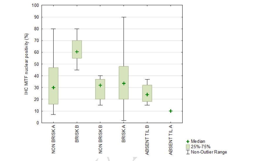
Melanoma progression is influenced by complex interactions between tumor cells and the immune microenvironment. This study examined the relationship between microphthalmia-associated transcription factor (MITF) expression and the immune microenvironment in primary melanoma using a modified classification of tumor-infiltrating lymphocytes (TILs) based on conventional BRISK categories. Archival formalin-fixed, paraffin-embedded tissue samples from 81 primary melanoma patients were analyzed via tissue microarray immunohistochemistry to assess MITF protein levels. TIL patterns were categorized into six groups, refining the traditional BRISK classification to distinguish between continuous and discontinuous infiltration, as well as peripheral vs intratumoral distribution. The analysis revealed that melanomas classified under the BRISK B category exhibited the highest MITF expression, often exceeding 50%. In contrast, tumors in the NON-BRISK and ABSENT TIL groups showed significantly lower MITF expression (mean values: 32.73% ± 16.98% and 22.00% ± 10.54%, respectively), with statistically significant differences (Kruskal–Wallis test, P = 0.027; modified classification, P = 0.011). Additionally, the presence of CD20+ B lymphocytes correlated with increased MITF expression (P = 0.009). MITF gene amplification was detected in 29% of cases, though its association with protein expression showed only a trend (P = 0.058). These findings highlight the complex interplay between MITF expression and TIL distribution in melanoma, suggesting that refined TIL classification may offer deeper insights into tumor immunobiology and help predict responses to immunotherapy.
Citations
Downloads
Downloads
Published
License
Copyright (c) 2025 Damir Vučinić, Matea Lekić, Gordana Žauhar, Gordana Zamolo

This work is licensed under a Creative Commons Attribution 4.0 International License.