A deep learning model based on chest CT to predict benign and malignant breast masses and axillary lymph node metastasis

Authors

  • Jingxiang Sun Department of Radiology, The First Affiliated Hospital of Shandong First Medical University & Shandong Provincial Qianfoshan Hospital, Shandong, China; Postgraduate Department, Shandong First Medical University (Shandong Academy of Medical Sciences), Shandong, China https://orcid.org/0009-0003-9916-9585
  • Xiaoming Xi School of Computer Science and Technology, Shandong Jianzhu University, Shandong, China
  • Mengying Wang School of Computer Science and Technology, Shandong Jianzhu University, Shandong, China
  • Menghan Liu Department of Radiology, The First Affiliated Hospital of Shandong First Medical University & Shandong Provincial Qianfoshan Hospital, Shandong, China
  • Xiaodong Zhang Postgraduate Department, Shandong First Medical University (Shandong Academy of Medical Sciences), Shandong, China
  • Haiyan Qiu Department of Radiology, The First Affiliated Hospital of Shandong First Medical University & Shandong Provincial Qianfoshan Hospital, Shandong, China; Postgraduate Department, Shandong First Medical University (Shandong Academy of Medical Sciences), Shandong, China
  • Youxin Zhang Department of Radiology, The First Affiliated Hospital of Shandong First Medical University & Shandong Provincial Qianfoshan Hospital, Shandong, China; Department of Postgraduate, Shandong Second Medical University, Shandong, China
  • Taian Fu Department of Radiology, The First Affiliated Hospital of Shandong First Medical University & Shandong Provincial Qianfoshan Hospital, Shandong, China; Department of Postgraduate, Shandong Second Medical University, Shandong, China https://orcid.org/0009-0005-1409-6503
  • Yanan Du Department of Health Management, The First Affiliated Hospital of Shandong First Medical University & Shandong Provincial Qianfoshan Hospital, Shandong, China
  • Wanqing Ren Department of Radiology, Jinan Third People’s Hospital, Shandong, China
  • Dawei Wang Department of Health Management, The First Affiliated Hospital of Shandong First Medical University & Shandong Provincial Qianfoshan Hospital, Shandong, China
  • Guang Zhang Department of Health Management, The First Affiliated Hospital of Shandong First Medical University & Shandong Provincial Qianfoshan Hospital, Shandong, China; Shandong Engineering Research Center of Health Management, Jinan, China; Shandong Institute of Health Management, Jinan, China https://orcid.org/0009-0006-2640-6019

DOI:

https://doi.org/10.17305/bb.2025.12010

Keywords:

Axillary lymph node metastasis, breast cancer, breast mass, chest CT, deep learning

Abstract

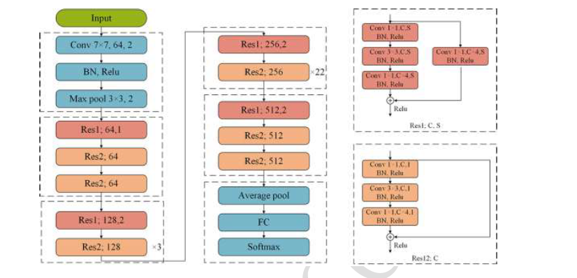
Differentiating early-stage breast cancer from benign breast masses is crucial for radiologists. Additionally, accurately assessing axillary lymph node metastasis (ALNM) plays a significant role in clinical management and prognosis for breast cancer patients. Chest computed tomography (CT) is a commonly used imaging modality in physical and preoperative evaluations. This study aims to develop a deep learning model based on chest CT imaging to improve the preliminary assessment of breast lesions, potentially reducing the need for costly follow-up procedures such as magnetic resonance imaging (MRI) or positron emission tomography-CT and alleviating the financial and emotional burden on patients. We retrospectively collected chest CT images from 482 patients with breast masses, classifying them as benign (n = 224) or malignant (n = 258) based on pathological findings. The malignant group was further categorized into ALNM-positive (n = 91) and ALNM-negative (n = 167) subgroups. Patients were randomly divided into training, validation, and test sets in an 8:1:1 ratio, with the test set excluded from model development. All patients underwent non-contrast chest CT before surgery. After preprocessing the images through cropping, scaling, and standardization, we applied ResNet-34, ResNet-50, and ResNet-101 architectures to differentiate between benign and malignant masses and to assess ALNM. Model performance was evaluated using sensitivity, specificity, accuracy, receiver operating characteristic (ROC) curves, and the area under the curve (AUC). The ResNet models effectively distinguished benign from malignant masses, with ResNet-101 achieving the highest performance (AUC: 0.964; 95% CI: 0.948–0.981). It also demonstrated excellent predictive capability for ALNM (AUC: 0.951; 95% CI: 0.926–0.975). In conclusion, these deep learning models show strong diagnostic potential for both breast mass classification and ALNM prediction, offering a valuable tool for improving clinical decision-making.

Citations

Downloads

Download data is not yet available.
A deep learning model based on chest CT to predict benign and malignant breast masses and axillary lymph node metastasis

Published

17-03-2025

Issue

Section

Thematic issue: AI/ML in diseases

Categories

How to Cite

1.
A deep learning model based on chest CT to predict benign and malignant breast masses and axillary lymph node metastasis. Biomol Biomed [Internet]. 2025 Mar. 17 [cited 2025 Mar. 22];. Available from: https://www.bjbms.org/ojs/index.php/bjbms/article/view/12010