Nogo-A exacerbates sepsis-associated encephalopathy by modulating microglial SHP-2/NLRP3 balance and inducing ROS and M1 polarization
DOI:
https://doi.org/10.17305/bb.2024.10822Keywords:
Nogo protein, encephalopathy, sepsis, endoplasmic reticulum stress, protein-tyrosine phosphatases, NLRP3 inflammasome, microglia, mitochondria, autophagy, apoptosisAbstract
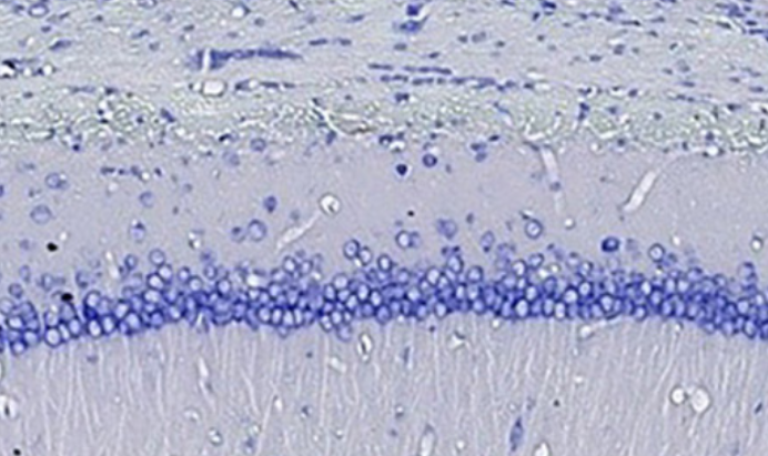
Sepsis, a systemic inflammatory response caused by infection, can lead to sepsis-associated encephalopathy (SAE), characterized by brain dysfunction without direct central nervous system infection. The pathogenesis of SAE involves blood-brain barrier disruption, neuroinflammation and neuronal death, with neuroinflammation being the core process. Nogo-A, a neurite growth-inhibitory protein in the central nervous system, is not well understood in sepsis. This study explores Nogo-A's mechanisms in sepsis, focusing on SAE. Using in vivo and in vitro methods, healthy SPF C57BL/6J male mice were divided into Sham, Nogo-A-NC-Model, and Nogo-A-KD-Model groups, with sepsis induced by abdominal ligation and puncture. Morris water maze tests assessed learning and memory, and brain tissues underwent hematoxylin-eosin (HE) staining, Nissl staining, and Western blot analysis. In vitro, Nogo-A gene knockdown models were constructed using BV-2 microglia cells to study inflammation and oxidative stress. Results showed Nogo-A expression affected learning and memory in septic mice, with knockdown reducing neuronal damage. Bioinformatics analysis suggested Nogo-A may activate reactive oxygen species (ROS) to inhibit p-SHP2, activating mitochondrial autophagy and promoting neuronal apoptosis. Western blot results confirmed that Nogo-A affects mitochondrial autophagy and neuronal survival by inhibiting SHP2 and activating ROS. Nogo-A's role in neuroinflammation and neuroprotection was emphasized, revealing its impact on endoplasmic reticulum (ER) stress, mitochondrial autophagy, and NLRP3 inflammasome activation. This study provides a theoretical basis for SAE treatment, suggesting further multi-gene and multi-pathway analyses and validation in clinical samples. Developing gene therapy and drug interventions targeting Nogo-A pathways will offer more effective treatment strategies.
Citations
Downloads
Downloads
Additional Files
Published
Issue
Section
Categories
License
Copyright (c) 2024 Ying Liu, Lei Guo, Guoan Zhang, Wenjie Sun, Xiaohui Yang, Yingfu Liu

This work is licensed under a Creative Commons Attribution 4.0 International License.


