Neuroimaging in the diagnosis and treatment of cerebral toxoplasmosis in children with severe β-thalassemia after allo-HSCT
DOI:
https://doi.org/10.17305/bb.2024.10708Keywords:
Cerebral toxoplasmosis, β-thalassemia major, allogeneic hematopoetic stem cell transplantation, magnetic resonance imaging, fluorodeoxyglucose positron emission tomography/computed tomography, metagenomic next generation sequencingAbstract
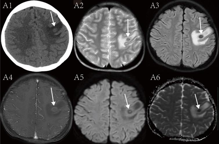
Children with severe β-thalassemia major (β-TM) are at high risk of developing toxoplasmosis after allogeneic hematopoietic stem cell transplantation (allo-HSCT). The aim of this study was to identify the neuroimaging findings of cerebral toxoplasmosis in pediatric patients with β-TM for early diagnosis and treatment of cerebral toxoplasmosis. We performed a retrospective assessment of clinical and neuroimaging data of children with severe β-TM who had cerebral toxoplasmosis after allo-HSCT. Additionally, we reviewed and summarized the literature on cerebral toxoplasmosis in patients with other underlying conditions. This case series identified three children who had severe β-TM and had subsequent cerebral toxoplasmosis after allo-HSCT. In addition, we identified 23 patients from literature who had toxoplasmosis and had underlying conditions other than β-TM. We found that the most common clinical symptom among the patients from our series and the patients from literature was fever upon presentation. We identified the typical neuroimaging findings including brain lesions with ring enhancement and eccentric/central nuclear target-like enhancement, which should facilitate early diagnosis and treatment of cerebral toxoplasmosis.
Citations
Downloads
Downloads
Additional Files
Published
License
Copyright (c) 2024 Meiai Liao, Guangrui Lai, Meiru Bu, Meiqing Wu, Muliang Jiang, Bihong T. Chen

This work is licensed under a Creative Commons Attribution 4.0 International License.