Feasibility and accuracy of a voxel-based neuronavigation system with 3D image rendering in preoperative planning and as a learning tool for young neurosurgeons, exemplified by the anatomical localization of the superior sagittal sinus

Authors

  • Guenther C. Feigl Department of Neurosurgery, General Hospital Klinikum Bamberg, Bamberg, Germany Department of Neurosurgery, University of Tuebingen Medical Center, Tuebingen, Germany https://orcid.org/0000-0001-8011-8997
  • Firas Thaher Department of Neurosurgery, Katharinenhospital, Klinikum Stuttgart, Stuttgart, Germany
  • Sören Danz Department of Neuroradiology, University of Tuebingen Medical Center, Tuebingen, Germany
  • Marcos Tatagiba Department of Neurosurgery, University of Tuebingen Medical Center, Tuebingen, Germany
  • Anne K. Hickmann Department of Neurosurgery, Katharinenhospital, Klinikum Stuttgart, Stuttgart, Germany https://orcid.org/0000-0002-6283-4348
  • Antje Fahrig Department of Radiotherapy and Radiooncology, General Hospital Klinikum Bamberg, Bamberg, Germany https://orcid.org/0000-0002-6283-4348
  • Tomaz Velnar Department of Neurosurgery, University Medical Centre Ljubljana, Ljubljana, Slovenia Alma Mater Europaea – European Center Maribor (AMEU-ECM), Maribor, Slovenia https://orcid.org/0000-0002-6283-4348
  • Marcel Kullmann Department of Neurosurgery, University of Tuebingen Medical Center, Tuebingen, Germany

DOI:

https://doi.org/10.17305/bjbms.2019.3815

Keywords:

Neuronavigation system, 3D volumetric image rendering, resident training, preoperative planning, anatomical relationship, superior sagittal sinus, sagittal suture, voxel-based

Abstract

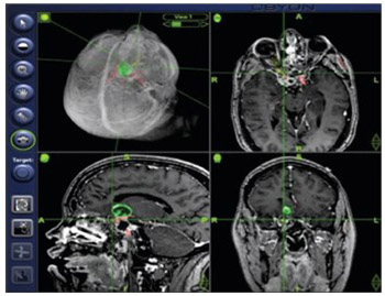
It is essential for a neurosurgeon to know individual anatomy and the corresponding anatomical landmarks before starting a surgery. Continuous training, especially of young neurosurgeons, is crucial for understanding complex neuroanatomy. In this study, we used a neuronavigation system with 3D volumetric image rendering to determine the anatomical relationship between the sagittal suture and the superior sagittal sinus (SSS) in patients with intracranial lesions. Furthermore, we discussed the applicability of such system in preoperative planning, residency training, and research. The study included 30 adult patients (18 female/12 male) who underwent a cranial computed tomography (CT) scan combined with venous angiography, for preoperative planning. The position of the sagittal suture in relation to the SSS was assessed in 3D CT images using an image guidance system (IGS) with 3D volumetric image rendering. Measurements were performed along the course of the sagittal sinus at the bregma, lambda, and in the middle between these two points. The SSS deviated to the right side of the sagittal suture in 50% of cases at the bregma, and in 46.7% at the midpoint and lambda. The SSS was displaced to the left of the sagittal suture in 10% of cases at the bregma and lambda and in 13% at the midpoint. IGSs with 3D volumetric image rendering enable simultaneous visualization of bony surfaces, soft tissue and vascular structures and interactive modulation of tissue transparency. They can be used in preoperative planning and intraoperative guidance to validate external landmarks and to determine anatomical relationships. In addition, 3D IGSs can be utilized for training of surgical residents and for research in anatomy.

Author Biographies

  • Guenther C. Feigl, Department of Neurosurgery, General Hospital Klinikum Bamberg, Bamberg, Germany Department of Neurosurgery, University of Tuebingen Medical Center, Tuebingen, Germany
    Department of Neurosurgery
  • Firas Thaher, Department of Neurosurgery, Katharinenhospital, Klinikum Stuttgart, Stuttgart, Germany
    Department of Neurosurgery
  • Sören Danz, Department of Neuroradiology, University of Tuebingen Medical Center, Tuebingen, Germany
    Department of Neuroradiology
  • Marcos Tatagiba, Department of Neurosurgery, University of Tuebingen Medical Center, Tuebingen, Germany
    Department of Neurosurgery
  • Anne K. Hickmann, Department of Neurosurgery, Katharinenhospital, Klinikum Stuttgart, Stuttgart, Germany
    Department of Neurosurgery
  • Antje Fahrig, Department of Radiotherapy and Radiooncology, General Hospital Klinikum Bamberg, Bamberg, Germany
    Department of Radiotherapy and Radiooncology
  • Tomaz Velnar, Department of Neurosurgery, University Medical Centre Ljubljana, Ljubljana, Slovenia Alma Mater Europaea – European Center Maribor (AMEU-ECM), Maribor, Slovenia
    Department of Neurosurgery
  • Marcel Kullmann, Department of Neurosurgery, University of Tuebingen Medical Center, Tuebingen, Germany
    Department of Neurosurgery
Feasibility and accuracy of a voxel-based neuronavigation system with 3D image rendering in preoperative planning and as a learning tool for young neurosurgeons, exemplified by the anatomical localization of the superior sagittal sinus

Downloads

Additional Files

Published

20-05-2019

Issue

Section

Translational and Clinical Research

Categories

How to Cite

1.
Feasibility and accuracy of a voxel-based neuronavigation system with 3D image rendering in preoperative planning and as a learning tool for young neurosurgeons, exemplified by the anatomical localization of the superior sagittal sinus. Biomol Biomed [Internet]. 2019 May 20 [cited 2024 Mar. 19];19(2):180-5. Available from: https://bjbms.org/ojs/index.php/bjbms/article/view/3815